 Movie
Movie Controller

[English] 日本語
 Yorodumi
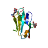
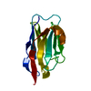
- PDB-5yyz: Crystal structure of the MEK1 FHA domain in complex with the HOP1... -
+ Open data

- Basic information
| Entry | Database: PDB / ID: 5yyz | ||||||
|---|---|---|---|---|---|---|---|
| Title | Crystal structure of the MEK1 FHA domain in complex with the HOP1 pThr318 peptide. | ||||||
 Components Components |
| ||||||
 Keywords Keywords | TRANSFERASE / MEK1 / HOP1 / Meiosis | ||||||
| Function / homology |  Function and homology information Function and homology informationactivation of reciprocal meiotic recombination / meiotic recombination checkpoint signaling / synaptonemal complex assembly / homologous chromosome pairing at meiosis / synaptonemal complex / homologous recombination / lateral element / reciprocal meiotic recombination / four-way junction DNA binding / condensed nuclear chromosome ...activation of reciprocal meiotic recombination / meiotic recombination checkpoint signaling / synaptonemal complex assembly / homologous chromosome pairing at meiosis / synaptonemal complex / homologous recombination / lateral element / reciprocal meiotic recombination / four-way junction DNA binding / condensed nuclear chromosome / meiotic cell cycle / protein kinase activity / non-specific serine/threonine protein kinase / protein serine kinase activity / protein serine/threonine kinase activity / chromatin binding / signal transduction / ATP binding / metal ion binding / nucleus / cytoplasm Similarity search - Function | ||||||
| Biological species |  | ||||||
| Method |  X-RAY DIFFRACTION / SYNCHROTRON / MOLECULAR REPLACEMENT / Resolution: 1.798 Å X-RAY DIFFRACTION / SYNCHROTRON / MOLECULAR REPLACEMENT / Resolution: 1.798 Å | ||||||
 Authors Authors | Xie, C. / Li, F. / Jiang, Y. / Wu, J. / Shi, Y. | ||||||
 Citation Citation | Journal: Acta Crystallogr D Struct Biol / Year: 2018 Title: Structural insights into the recognition of phosphorylated Hop1 by Mek1 Authors: Xie, C. / He, C. / Jiang, Y. / Yu, H. / Cheng, L. / Nshogoza, G. / Ala, M.S. / Tian, C. / Wu, J. / Shi, Y. / Li, F. | ||||||
| History |
|
- Structure visualization
| Structure viewer | Molecule: MolmilJmol/JSmol |
|---|
- Downloads & links
-Download
| PDBx/mmCIF format | 5yyz.cif.gz | 65.4 KB | Display | PDBx/mmCIF format |
|---|---|---|---|---|
| PDB format | pdb5yyz.ent.gz | 46.1 KB | Display | PDB format |
| PDBx/mmJSON format | 5yyz.json.gz | Tree view | PDBx/mmJSON format | |
| Others |  Other downloads Other downloads |
-Validation report
| Arichive directory | https://data.pdbj.org/pub/pdb/validation_reports/yy/5yyzftp://data.pdbj.org/pub/pdb/validation_reports/yy/5yyz | HTTPS FTP |
|---|
-Related structure data
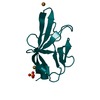
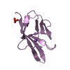
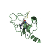
| Related structure data |  5yyxC  1g6gS S: Starting model for refinement C: citing same article ( |
|---|---|
| Similar structure data |


-Links
 PDBj
PDBj


- Assembly
| Deposited unit | 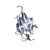
| ||||||||
|---|---|---|---|---|---|---|---|---|---|
| 1 | 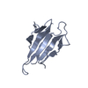
| ||||||||
| 2 | 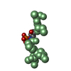
| ||||||||
| Unit cell |
|
-Components
| #1: Protein | Mass: 16107.298 Da / Num. of mol.: 1 Source method: isolated from a genetically manipulated source Source: (gene. exp.) Production host:  References: UniProt: P24719, non-specific serine/threonine protein kinase |
|---|---|
| #2: Protein/peptide | Mass: 1513.502 Da / Num. of mol.: 1 / Source method: obtained synthetically / Source: (synth.) |
| #3: Water | ChemComp-HOH /  Mass: 18.015 Da / Num. of mol.: 107 / Source method: isolated from a natural source / Formula: H2O Mass: 18.015 Da / Num. of mol.: 107 / Source method: isolated from a natural source / Formula: H2O |
| Has protein modification | Y |
-Experimental details
-Experiment
| Experiment | Method: X-RAY DIFFRACTION / Number of used crystals: 1 |
|---|
- Sample preparation
| Crystal | Density Matthews: 2.37 Å3/Da / Density % sol: 48.06 % |
|---|---|
| Crystal grow | Temperature: 293 K / Method: vapor diffusion, hanging drop Details: 0.2M Lithium Citrate tribasic tetrahydrate, 20% w/v PEG 3350 |
-Data collection
| Diffraction | Mean temperature: 100 K | |||||||||||||||||||||||||||||||||||||||||||||||||||||||||||||||||||||||||||||||||||||||||||||||||||
|---|---|---|---|---|---|---|---|---|---|---|---|---|---|---|---|---|---|---|---|---|---|---|---|---|---|---|---|---|---|---|---|---|---|---|---|---|---|---|---|---|---|---|---|---|---|---|---|---|---|---|---|---|---|---|---|---|---|---|---|---|---|---|---|---|---|---|---|---|---|---|---|---|---|---|---|---|---|---|---|---|---|---|---|---|---|---|---|---|---|---|---|---|---|---|---|---|---|---|---|---|
| Diffraction source | Source: SYNCHROTRON / Site: SSRF  / Beamline: BL19U1 / Wavelength: 0.97853 Å / Beamline: BL19U1 / Wavelength: 0.97853 Å | |||||||||||||||||||||||||||||||||||||||||||||||||||||||||||||||||||||||||||||||||||||||||||||||||||
| Detector | Type: DECTRIS PILATUS 6M / Detector: PIXEL / Date: Dec 22, 2016 | |||||||||||||||||||||||||||||||||||||||||||||||||||||||||||||||||||||||||||||||||||||||||||||||||||
| Radiation | Protocol: SINGLE WAVELENGTH / Monochromatic (M) / Laue (L): M / Scattering type: x-ray | |||||||||||||||||||||||||||||||||||||||||||||||||||||||||||||||||||||||||||||||||||||||||||||||||||
| Radiation wavelength | Wavelength: 0.97853 Å / Relative weight: 1 | |||||||||||||||||||||||||||||||||||||||||||||||||||||||||||||||||||||||||||||||||||||||||||||||||||
| Reflection | Resolution: 1.798→40 Å / Num. obs: 15415 / % possible obs: 99.1 % / Redundancy: 6.6 % / Biso Wilson estimate: 26.38 Å2 / Rmerge(I) obs: 0.061 / Rpim(I) all: 0.025 / Rrim(I) all: 0.066 / Χ2: 0.525 / Net I/σ(I): 5.6 | |||||||||||||||||||||||||||||||||||||||||||||||||||||||||||||||||||||||||||||||||||||||||||||||||||
| Reflection shell | Diffraction-ID: 1
|
- Processing
| Software |
| ||||||||||||||||||||||||||||||||||||||||||
|---|---|---|---|---|---|---|---|---|---|---|---|---|---|---|---|---|---|---|---|---|---|---|---|---|---|---|---|---|---|---|---|---|---|---|---|---|---|---|---|---|---|---|---|
| Refinement | Method to determine structure: MOLECULAR REPLACEMENT Starting model: 1G6G Resolution: 1.798→33.701 Å / SU ML: 0.2 / Cross valid method: THROUGHOUT / σ(F): 1.39 / Phase error: 24.39 / Stereochemistry target values: ML
| ||||||||||||||||||||||||||||||||||||||||||
| Solvent computation | Shrinkage radii: 0.9 Å / VDW probe radii: 1.11 Å / Solvent model: FLAT BULK SOLVENT MODEL | ||||||||||||||||||||||||||||||||||||||||||
| Displacement parameters | Biso max: 105.76 Å2 / Biso mean: 36.3355 Å2 / Biso min: 14.97 Å2 | ||||||||||||||||||||||||||||||||||||||||||
| Refinement step | Cycle: final / Resolution: 1.798→33.701 Å
| ||||||||||||||||||||||||||||||||||||||||||
| Refine LS restraints |
| ||||||||||||||||||||||||||||||||||||||||||
| LS refinement shell | Refine-ID: X-RAY DIFFRACTION / Rfactor Rfree error: 0 / Total num. of bins used: 5
| ||||||||||||||||||||||||||||||||||||||||||
| Refinement TLS params. | Method: refined / Origin x: 27.1255 Å / Origin y: 12.7781 Å / Origin z: 33.1246 Å
| ||||||||||||||||||||||||||||||||||||||||||
| Refinement TLS group |
|
