 Movie
Movie Controller

[English] 日本語
 Yorodumi
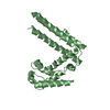
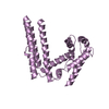
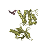
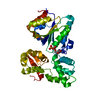
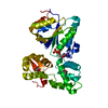
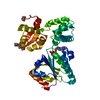
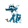
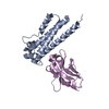
- PDB-4iap: Crystal structure of PH domain of Osh3 from Saccharomyces cerevisiae -
+ Open data

- Basic information
| Entry | Database: PDB / ID: 4iap | ||||||
|---|---|---|---|---|---|---|---|
| Title | Crystal structure of PH domain of Osh3 from Saccharomyces cerevisiae | ||||||
 Components Components | Oxysterol-binding protein homolog 3,Endolysin,Oxysterol-binding protein homolog 3 | ||||||
 Keywords Keywords | LIPID BINDING PROTEIN/ HYDRORASE / PH domain / beta sandwitch / targeting / phosphoinositides / LIPID BINDING PROTEIN- HYDRORASE complex | ||||||
| Function / homology |  Function and homology information Function and homology informationSynthesis of bile acids and bile salts / karyogamy involved in conjugation with cellular fusion / ER to Golgi ceramide transport / sterol transfer activity / pseudohyphal growth / invasive growth in response to glucose limitation / sterol binding / sterol transport / perinuclear endoplasmic reticulum / cortical endoplasmic reticulum ...Synthesis of bile acids and bile salts / karyogamy involved in conjugation with cellular fusion / ER to Golgi ceramide transport / sterol transfer activity / pseudohyphal growth / invasive growth in response to glucose limitation / sterol binding / sterol transport / perinuclear endoplasmic reticulum / cortical endoplasmic reticulum / maintenance of cell polarity / piecemeal microautophagy of the nucleus / reticulophagy / exocytosis / viral release from host cell by cytolysis / peptidoglycan catabolic process / cell wall macromolecule catabolic process / endocytosis / lysozyme / lysozyme activity / host cell cytoplasm / defense response to bacterium / lipid binding / endoplasmic reticulum membrane / plasma membrane / cytosol / cytoplasm Similarity search - Function | ||||||
| Biological species |   Enterobacteria phage T4 (virus) Enterobacteria phage T4 (virus) | ||||||
| Method |  X-RAY DIFFRACTION / SYNCHROTRON / MOLECULAR REPLACEMENT / Resolution: 2.3 Å X-RAY DIFFRACTION / SYNCHROTRON / MOLECULAR REPLACEMENT / Resolution: 2.3 Å | ||||||
 Authors Authors | Tong, J. / Im, Y.J. | ||||||
 Citation Citation | Journal: Structure / Year: 2013 Title: Structure of osh3 reveals a conserved mode of phosphoinositide binding in oxysterol-binding proteins Authors: Tong, J. / Yang, H. / Yang, H. / Eom, S.H. / Im, Y.J. | ||||||
| History |
|
- Structure visualization
| Structure viewer | Molecule: MolmilJmol/JSmol |
|---|
- Downloads & links
-Download
| PDBx/mmCIF format | 4iap.cif.gz | 116.3 KB | Display | PDBx/mmCIF format |
|---|---|---|---|---|
| PDB format | pdb4iap.ent.gz | 90.4 KB | Display | PDB format |
| PDBx/mmJSON format | 4iap.json.gz | Tree view | PDBx/mmJSON format | |
| Others |  Other downloads Other downloads |
-Validation report
| Arichive directory | https://data.pdbj.org/pub/pdb/validation_reports/ia/4iapftp://data.pdbj.org/pub/pdb/validation_reports/ia/4iap | HTTPS FTP |
|---|
-Related structure data
| Related structure data |  4ic4C  4inqC  212lS S: Starting model for refinement C: citing same article ( |
|---|---|
| Similar structure data |


-Links
 PDBj
PDBj


- Assembly
| Deposited unit | 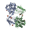
| ||||||||
|---|---|---|---|---|---|---|---|---|---|
| 1 | 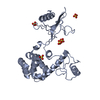
| ||||||||
| 2 | 
| ||||||||
| 3 |
| ||||||||
| Unit cell |
|
-Components
| #1: Protein | Mass: 29746.031 Da / Num. of mol.: 2 Fragment: PH domain (UNP residues 221-317), T4 Lysozyme (UNP residues 2-161),PH domain (UNP residues 237-315) Mutation: D1020N, C1054T, C1097A Source method: isolated from a genetically manipulated source Source: (gene. exp.) Enterobacteria phage T4 (virus)Strain: ATCC 204508 / S288c / Gene: OSH3, YHR073W / Plasmid: modifed pHIS / Production host:  #2: Chemical | ChemComp-SO4 /   Mass: 96.063 Da / Num. of mol.: 8 / Source method: obtained synthetically / Formula: SO4 Mass: 96.063 Da / Num. of mol.: 8 / Source method: obtained synthetically / Formula: SO4#3: Water | ChemComp-HOH / |  Mass: 18.015 Da / Num. of mol.: 103 / Source method: isolated from a natural source / Formula: H2O Mass: 18.015 Da / Num. of mol.: 103 / Source method: isolated from a natural source / Formula: H2OSequence details | THERE ARE SEQUENCE CONFLICTS IN THE UNP SEQUENCE REFERENCE P00720. | |
|---|
-Experimental details
-Experiment
| Experiment | Method: X-RAY DIFFRACTION / Number of used crystals: 1 |
|---|
- Sample preparation
| Crystal | Density Matthews: 3.13 Å3/Da / Density % sol: 60.69 % |
|---|---|
| Crystal grow | Temperature: 295 K / Method: vapor diffusion, hanging drop / pH: 8 Details: 0.1M Tris-HCl, 15% PEG8000, 0.2 M Li2SO4, pH 8.0, VAPOR DIFFUSION, HANGING DROP, temperature 295K |
-Data collection
| Diffraction | Mean temperature: 100 K |
|---|---|
| Diffraction source | Source: SYNCHROTRON / Site: PAL/PLS  / Beamline: 4A / Wavelength: 0.97949 Å / Beamline: 4A / Wavelength: 0.97949 Å |
| Detector | Type: ADSC QUANTUM 315 / Detector: CCD / Date: Jul 23, 2012 / Details: mirrors |
| Radiation | Monochromator: Si 111 CHANNEL / Protocol: SINGLE WAVELENGTH / Monochromatic (M) / Laue (L): M / Scattering type: x-ray |
| Radiation wavelength | Wavelength: 0.97949 Å / Relative weight: 1 |
| Reflection | Resolution: 2.3→50 Å / Num. all: 33493 / Num. obs: 32983 / % possible obs: 99.4 % / Observed criterion σ(F): 3 / Observed criterion σ(I): 3 / Redundancy: 3.3 % / Biso Wilson estimate: 29.9 Å2 / Rmerge(I) obs: 0.067 / Net I/σ(I): 23.2 |
| Reflection shell | Resolution: 2.3→2.34 Å / Redundancy: 3 % / Rmerge(I) obs: 0.308 / Mean I/σ(I) obs: 3.5 / Num. unique all: 1666 / % possible all: 98.7 |
- Processing
| Software |
| ||||||||||||||||||||||||||||||||||||||||||||||||||||||||||||
|---|---|---|---|---|---|---|---|---|---|---|---|---|---|---|---|---|---|---|---|---|---|---|---|---|---|---|---|---|---|---|---|---|---|---|---|---|---|---|---|---|---|---|---|---|---|---|---|---|---|---|---|---|---|---|---|---|---|---|---|---|---|
| Refinement | Method to determine structure: MOLECULAR REPLACEMENT Starting model: T4 Lysozyme 212L Resolution: 2.3→40.02 Å / Rfactor Rfree error: 0.007 / Data cutoff high absF: 1788889.68 / Data cutoff low absF: 0 / Isotropic thermal model: RESTRAINED / Cross valid method: THROUGHOUT / σ(F): 0 / Stereochemistry target values: Engh & Huber
| ||||||||||||||||||||||||||||||||||||||||||||||||||||||||||||
| Solvent computation | Solvent model: FLAT MODEL / Bsol: 31.8257 Å2 / ksol: 0.328213 e/Å3 | ||||||||||||||||||||||||||||||||||||||||||||||||||||||||||||
| Displacement parameters | Biso mean: 43.6 Å2
| ||||||||||||||||||||||||||||||||||||||||||||||||||||||||||||
| Refine analyze |
| ||||||||||||||||||||||||||||||||||||||||||||||||||||||||||||
| Refinement step | Cycle: LAST / Resolution: 2.3→40.02 Å
| ||||||||||||||||||||||||||||||||||||||||||||||||||||||||||||
| Refine LS restraints |
| ||||||||||||||||||||||||||||||||||||||||||||||||||||||||||||
| LS refinement shell | Resolution: 2.3→2.44 Å / Rfactor Rfree error: 0.02 / Total num. of bins used: 6
| ||||||||||||||||||||||||||||||||||||||||||||||||||||||||||||
| Xplor file |
|
