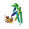
Entry Database : PDB / ID : 6d8zTitle Crystal Structure of the C-terminal Guanine Nucleotide Exchange Factor Module of Human Trio Triple functional domain protein Keywords / / / / Function / homology Function Domain/homology Component
/ / / / / / / / / / / / / / / / / / / / / / / / / / / / / / / / / / / / / / / / / / / / / / / / / / / / / / / / / / / / / / / / / / / / / / / / / / / / / / / / / / / / / / / / / / / / / / / / / / / / / / / / Biological species Homo sapiens (human)Method / / / Resolution : 2.65 Å Authors Bandekar, S. / Tesmer, J.J. Funding support Organization Grant number Country National Institutes of Health/National Heart, Lung, and Blood Institute (NIH/NHLBI) R01 HL122416-02z National Institutes of Health/National Heart, Lung, and Blood Institute (NIH/NHLBI) R01 HL071818 National Institutes of Health/National Cancer Institute (NIH/NCI) R01 CA221289
Journal : Sci Signal / Year : 2019Title : Structure of the C-terminal guanine nucleotide exchange factor module of Trio in an autoinhibited conformation reveals its oncogenic potential.Authors : Bandekar, S.J. / Arang, N. / Tully, E.S. / Tang, B.A. / Barton, B.L. / Li, S. / Gutkind, J.S. / Tesmer, J.J.G. History Deposition Apr 27, 2018 Deposition site / Processing site Revision 1.0 Feb 20, 2019 Provider / Type Revision 1.1 Feb 27, 2019 Group / Database references / Category / Item / _citation.titleRevision 1.2 Mar 6, 2019 Group / Database referencesCategory citation / citation_author ... citation / citation_author / database_PDB_rev / database_PDB_rev_record / pdbx_database_proc Item _citation.journal_abbrev / _citation.journal_volume ... _citation.journal_abbrev / _citation.journal_volume / _citation.pdbx_database_id_PubMed / _citation.title Revision 1.3 Dec 4, 2019 Group / Category / Item Revision 1.4 May 25, 2022 Group / Structure summary / Category / structItem / _database_2.pdbx_database_accession / _struct.titleRevision 1.5 Oct 4, 2023 Group / Refinement descriptionCategory / chem_comp_bond / pdbx_initial_refinement_model
Show all Show less
 Movie
Movie Controller

 Yorodumi
 Open data
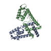
 Basic information
 Components
 Keywords
 Function and homology information
 Homo sapiens (human)
 X-RAY DIFFRACTION /
  Authors
 United States, 3items
  Citation
 Structure visualization
 Downloads & links
 Other downloads
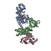


 PDBj
PDBj


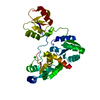
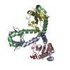
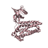
 Assembly
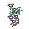


 Mass: 18.015 Da / Num. of mol.: 26 / Source method: isolated from a natural source / Formula: H2O
 Sample preparation
 Processing