 Movie
Movie Controller

[English] 日本語
 Yorodumi
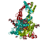
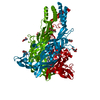
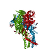
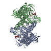
- PDB-4dw1: Crystal structure of the ATP-gated P2X4 ion channel in the ATP-bo... -
+ Open data

- Basic information
| Entry | Database: PDB / ID: 4dw1 | |||||||||
|---|---|---|---|---|---|---|---|---|---|---|
| Title | Crystal structure of the ATP-gated P2X4 ion channel in the ATP-bound, open state at 2.8 Angstroms | |||||||||
 Components Components | P2X purinoceptor | |||||||||
 Keywords Keywords | TRANSPORT PROTEIN / ion channel | |||||||||
| Function / homology |  Function and homology information Function and homology informationElevation of cytosolic Ca2+ levels / Platelet homeostasis / purine nucleotide binding / extracellularly ATP-gated monoatomic cation channel activity / purinergic nucleotide receptor activity / ATP-gated ion channel activity / transmembrane transporter complex / CTP binding / response to ATP / monoatomic ion channel complex ...Elevation of cytosolic Ca2+ levels / Platelet homeostasis / purine nucleotide binding / extracellularly ATP-gated monoatomic cation channel activity / purinergic nucleotide receptor activity / ATP-gated ion channel activity / transmembrane transporter complex / CTP binding / response to ATP / monoatomic ion channel complex / monoatomic cation transport / ligand-gated monoatomic ion channel activity / calcium ion transport / postsynapse / lysosomal membrane / ATP binding / membrane / identical protein binding / plasma membrane Similarity search - Function | |||||||||
| Biological species |  | |||||||||
| Method |  X-RAY DIFFRACTION / SYNCHROTRON / MOLECULAR REPLACEMENT / Resolution: 2.8 Å X-RAY DIFFRACTION / SYNCHROTRON / MOLECULAR REPLACEMENT / Resolution: 2.8 Å | |||||||||
 Authors Authors | Hattori, M. / Gouaux, E. | |||||||||
 Citation Citation | Journal: Nature / Year: 2012 Title: Molecular mechanism of ATP binding and ion channel activation in P2X receptors. Authors: Hattori, M. / Gouaux, E. | |||||||||
| History |
|
- Structure visualization
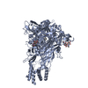
| Structure viewer | Molecule: MolmilJmol/JSmol |
|---|
- Downloads & links
-Download
| PDBx/mmCIF format | 4dw1.cif.gz | 85.6 KB | Display | PDBx/mmCIF format |
|---|---|---|---|---|
| PDB format | pdb4dw1.ent.gz | 61.7 KB | Display | PDB format |
| PDBx/mmJSON format | 4dw1.json.gz | Tree view | PDBx/mmJSON format | |
| Others |  Other downloads Other downloads |
-Validation report
| Arichive directory | https://data.pdbj.org/pub/pdb/validation_reports/dw/4dw1ftp://data.pdbj.org/pub/pdb/validation_reports/dw/4dw1 | HTTPS FTP |
|---|
-Related structure data


-Links
 PDBj
PDBj- Assembly
| Deposited unit | 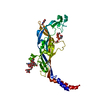
| ||||||||
|---|---|---|---|---|---|---|---|---|---|
| 1 | 
| ||||||||
| Unit cell |
| ||||||||
| Details | The biological assembly is a trimer generated from the protomer in the asymmetric unit by the operations: -y, x-y, z and y-x, -x, z. |
-Components
-Protein , 1 types, 1 molecules A
| #1: Protein | Mass: 38117.625 Da / Num. of mol.: 1 / Fragment: UNP residues 28-365 / Mutation: N78K, N187R, H252R Source method: isolated from a genetically manipulated source Source: (gene. exp.)   Spodoptera frugiperda (fall armyworm) / Strain (production host): Sf9 / References: UniProt: Q6NYR1, UniProt: F8W463*PLUS Spodoptera frugiperda (fall armyworm) / Strain (production host): Sf9 / References: UniProt: Q6NYR1, UniProt: F8W463*PLUS |
|---|
-Sugars , 2 types, 2 molecules 
| #2: Polysaccharide | alpha-D-mannopyranose-(1-3)-alpha-D-mannopyranose-(1-3)-[alpha-D-mannopyranose-(1-6)]beta-D- ...alpha-D-mannopyranose-(1-3)-alpha-D-mannopyranose-(1-3)-[alpha-D-mannopyranose-(1-6)]beta-D-mannopyranose-(1-4)-2-acetamido-2-deoxy-beta-D-glucopyranose-(1-4)-2-acetamido-2-deoxy-beta-D-glucopyranose Source method: isolated from a genetically manipulated source |
|---|---|
| #3: Sugar | ChemComp-NAG /  Type: D-saccharide, beta linking / Mass: 221.208 Da / Num. of mol.: 1 Type: D-saccharide, beta linking / Mass: 221.208 Da / Num. of mol.: 1Source method: isolated from a genetically manipulated source Formula: C8H15NO6 |
-Non-polymers , 3 types, 95 molecules 


| #4: Chemical | ChemComp-ATP /  Mass: 507.181 Da / Num. of mol.: 1 / Source method: obtained synthetically / Formula: C10H16N5O13P3 / Comment: ATP, energy-carrying molecule*YM Mass: 507.181 Da / Num. of mol.: 1 / Source method: obtained synthetically / Formula: C10H16N5O13P3 / Comment: ATP, energy-carrying molecule*YM | ||
|---|---|---|---|
| #5: Chemical |  Mass: 92.094 Da / Num. of mol.: 2 / Source method: obtained synthetically / Formula: C3H8O3 Mass: 92.094 Da / Num. of mol.: 2 / Source method: obtained synthetically / Formula: C3H8O3#6: Water | ChemComp-HOH / | Mass: 18.015 Da / Num. of mol.: 92 / Source method: isolated from a natural source / Formula: H2O |
-Details
| Has protein modification | Y |
|---|
-Experimental details
-Experiment
| Experiment | Method: X-RAY DIFFRACTION / Number of used crystals: 5 |
|---|
- Sample preparation
| Crystal | Density Matthews: 5.28 Å3/Da / Density % sol: 76.69 % |
|---|---|
| Crystal grow | Temperature: 277 K / Method: vapor diffusion, hanging drop / pH: 8 Details: 1 mM ATP, 20-26% PEG 2000, 300 mM Mg(NO3)2, 100 mM Tris, pH 8, VAPOR DIFFUSION, HANGING DROP, temperature 277K |
-Data collection
| Diffraction | Mean temperature: 100 K |
|---|---|
| Diffraction source | Source: SYNCHROTRON / Site: APS  / Beamline: 24-ID-C / Wavelength: 0.97918 Å / Beamline: 24-ID-C / Wavelength: 0.97918 Å |
| Detector | Type: ADSC QUANTUM 315 / Detector: CCD / Date: Mar 28, 2011 |
| Radiation | Monochromator: Cryo-Cooled Si(111) double crystal / Protocol: SINGLE WAVELENGTH / Monochromatic (M) / Laue (L): M / Scattering type: x-ray |
| Radiation wavelength | Wavelength: 0.97918 Å / Relative weight: 1 |
| Reflection | Resolution: 2.8→50 Å / Num. all: 20144 / Num. obs: 19941 / % possible obs: 99 % / Observed criterion σ(I): -1 / Redundancy: 7.8 % |
| Reflection shell | Resolution: 2.8→2.85 Å / % possible all: 98.4 |
- Processing
| Software |
| ||||||||||||||||||||||||||||||||||||||||||||||||||||||||
|---|---|---|---|---|---|---|---|---|---|---|---|---|---|---|---|---|---|---|---|---|---|---|---|---|---|---|---|---|---|---|---|---|---|---|---|---|---|---|---|---|---|---|---|---|---|---|---|---|---|---|---|---|---|---|---|---|---|
| Refinement | Method to determine structure: MOLECULAR REPLACEMENT / Resolution: 2.8→45.94 Å / SU ML: 0.3 / σ(F): 0 / Phase error: 21.84 / Stereochemistry target values: ML
| ||||||||||||||||||||||||||||||||||||||||||||||||||||||||
| Solvent computation | Shrinkage radii: 0.95 Å / VDW probe radii: 1.2 Å / Solvent model: FLAT BULK SOLVENT MODEL / Bsol: 80.039 Å2 / ksol: 0.301 e/Å3 | ||||||||||||||||||||||||||||||||||||||||||||||||||||||||
| Displacement parameters |
| ||||||||||||||||||||||||||||||||||||||||||||||||||||||||
| Refinement step | Cycle: LAST / Resolution: 2.8→45.94 Å
| ||||||||||||||||||||||||||||||||||||||||||||||||||||||||
| Refine LS restraints |
| ||||||||||||||||||||||||||||||||||||||||||||||||||||||||
| LS refinement shell |
|
