[English] 日本語
Yorodumi
- PDB-4nmn: Aquifex aeolicus replicative helicase (DnaB) complexed with ADP, ... -

+
Open data


ID or keywords:

Loading...

-
Basic information

Entry
Database: PDB / ID: 4nmn
Title
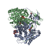
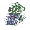
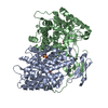
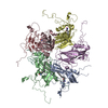
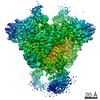
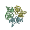
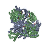
Aquifex aeolicus replicative helicase (DnaB) complexed with ADP, at 3.3 resolution
ComponentsReplicative DNA helicase
KeywordsREPLICATION / RecA-type helicase / Replicative DNA helicase / ATP binding / DNA-binding
Function / homology
Function and homology information


primosome complex / DNA replication, synthesis of RNA primer / DNA duplex unwinding / DNA unwinding involved in DNA replication / DNA helicase activity / DNA helicase / ATP hydrolysis activity / DNA binding / ATP binding / metal ion binding / cytosol
Similarity search - Function
DNA helicase, DnaB type / DNA helicase, DnaB-like, N-terminal / DnaB-like helicase N terminal domain / DNA helicase, DnaB-like, N-terminal domain superfamily / DNA helicase DnaB, N-terminal/DNA primase DnaG, C-terminal / DnaB-like helicase C terminal domain / DNA helicase, DnaB-like, C-terminal / Superfamily 4 helicase domain profile. / P-loop containing nucleotide triphosphate hydrolases / ATPases associated with a variety of cellular activities ...DNA helicase, DnaB type / DNA helicase, DnaB-like, N-terminal / DnaB-like helicase N terminal domain / DNA helicase, DnaB-like, N-terminal domain superfamily / DNA helicase DnaB, N-terminal/DNA primase DnaG, C-terminal / DnaB-like helicase C terminal domain / DNA helicase, DnaB-like, C-terminal / Superfamily 4 helicase domain profile. / P-loop containing nucleotide triphosphate hydrolases / ATPases associated with a variety of cellular activities / AAA+ ATPase domain / P-loop containing nucleoside triphosphate hydrolase / Rossmann fold / 3-Layer(aba) Sandwich / Alpha Beta
Similarity search - Domain/homology
ADENOSINE-5'-DIPHOSPHATE / STRONTIUM ION / Replicative DNA helicase
Similarity search - Component
Biological speciesAquifex aeolicus (bacteria)
MethodX-RAY DIFFRACTION / SYNCHROTRON / MOLECULAR REPLACEMENT / Resolution: 3.301 Å
AuthorsLyubimov, A.Y. / Strycharska, M.S. / Erzberger, J.P. / Berger, J.M.
CitationJournal: Mol Cell / Year: 2013
Title: Nucleotide and partner-protein control of bacterial replicative helicase structure and function.
Authors: Melania S Strycharska / Ernesto Arias-Palomo / Artem Y Lyubimov / Jan P Erzberger / Valerie L O'Shea / Carlos J Bustamante / James M Berger /
Abstract: Cellular replication forks are powered by ring-shaped, hexameric helicases that encircle and unwind DNA. To better understand the molecular mechanisms and control of these enzymes, we used multiple ...Cellular replication forks are powered by ring-shaped, hexameric helicases that encircle and unwind DNA. To better understand the molecular mechanisms and control of these enzymes, we used multiple methods to investigate the bacterial replicative helicase, DnaB. A 3.3 Å crystal structure of Aquifex aeolicus DnaB, complexed with nucleotide, reveals a newly discovered conformational state for this motor protein. Electron microscopy and small angle X-ray scattering studies confirm the state seen crystallographically, showing that the DnaB ATPase domains and an associated N-terminal collar transition between two physical states in a nucleotide-dependent manner. Mutant helicases locked in either collar state are active but display different capacities to support critical activities such as duplex translocation and primase-dependent RNA synthesis. Our findings establish the DnaB collar as an autoregulatory hub that controls the ability of the helicase to transition between different functional states in response to both nucleotide and replication initiation/elongation factors.
History
DepositionNov 15, 2013Deposition site: RCSB / Processing site: RCSB
Revision 1.0Jan 29, 2014Provider: repository / Type: Initial release

-
Structure visualization

Structure viewerMolecule:
MolmilJmol/JSmol

Downloads & links

-
Assembly

Deposited unit
A: Replicative DNA helicase
B: Replicative DNA helicase
hetero molecules


Theoretical massNumber of molelcules
Total (without water)100,3877
Polymers99,3972
Non-polymers9915
Water27015
1
A: Replicative DNA helicase
B: Replicative DNA helicase
hetero molecules

A: Replicative DNA helicase
B: Replicative DNA helicase
hetero molecules

A: Replicative DNA helicase
B: Replicative DNA helicase
hetero molecules


Theoretical massNumber of molelcules
Total (without water)301,16121
Polymers298,1906
Non-polymers2,97215
Water1086
TypeNameSymmetry operationNumber
identity operation1_555x,y,z1
crystal symmetry operation5_555z,x,y1
crystal symmetry operation9_555y,z,x1
Buried area34260 Å2
ΔGint-149 kcal/mol
Surface area107350 Å2
MethodPISA
Unit cell
Length a, b, c (Å)195.707, 195.707, 195.707
Angle α, β, γ (deg.)90.00, 90.00, 90.00
Int Tables number199
Space group name H-MI213
Components on special symmetry positions
IDModelComponents
11A-1003-

SR

21A-1101-

HOH

31A-1109-

HOH

-
Components

#1: Protein Replicative DNA helicase


Mass: 49698.266 Da / Num. of mol.: 2 / Fragment: UNP residues 7-440
Source method: isolated from a genetically manipulated source
Source: (gene. exp.) Aquifex aeolicus (bacteria) / Strain: VF5 / Gene: dnaB, aq_1472 / Production host: Escherichia coli (E. coli) / References: UniProt: O67450
#2: Chemical ChemComp-ADP / ADENOSINE-5'-DIPHOSPHATE / Adenosine diphosphate


Mass: 427.201 Da / Num. of mol.: 2 / Source method: obtained synthetically / Formula: C10H15N5O10P2 / Comment: ADP, energy-carrying molecule*YM
#3: Chemical ChemComp-MG / MAGNESIUM ION


Mass: 24.305 Da / Num. of mol.: 2 / Source method: obtained synthetically / Formula: Mg
#4: Chemical ChemComp-SR / STRONTIUM ION / Strontium


Mass: 87.620 Da / Num. of mol.: 1 / Source method: obtained synthetically / Formula: Sr
#5: Water ChemComp-HOH / water / Water


Mass: 18.015 Da / Num. of mol.: 15 / Source method: isolated from a natural source / Formula: H2O

-
Experimental details

-
Experiment

ExperimentMethod: X-RAY DIFFRACTION / Number of used crystals: 1

-
Sample preparation

CrystalDensity Matthews: 3.15 Å3/Da / Density % sol: 60.93 %
Crystal growTemperature: 294 K / Method: vapor diffusion, hanging drop / pH: 7.5
Details: 100mM Hepes pH 7.5, 200-300mM KSCN or NaSCN and 18-22% PEG 3350, VAPOR DIFFUSION, HANGING DROP, temperature 294K

-
Data collection

DiffractionMean temperature: 100 K
Diffraction sourceSource: SYNCHROTRON / Site: ALS / Beamline: 8.3.1 / Wavelength: 1.1 Å
DetectorType: ADSC QUANTUM 210 / Detector: CCD
RadiationMonochromator: Double flat crystal, Si(111) / Protocol: SINGLE WAVELENGTH / Monochromatic (M) / Laue (L): M / Scattering type: x-ray
Radiation wavelengthWavelength: 1.1 Å / Relative weight: 1
ReflectionResolution: 3.3→69.2 Å / Num. all: 18616 / Num. obs: 18616 / % possible obs: 98.4 % / Observed criterion σ(F): 1.36 / Observed criterion σ(I): 1.36 / Redundancy: 5.5 % / Biso Wilson estimate: 92.7 Å2 / Rmerge(I) obs: 0.116 / Net I/σ(I): 8.9
Reflection shellResolution: 3.3→3.5 Å / Redundancy: 5.2 % / Rmerge(I) obs: 0.554 / Mean I/σ(I) obs: 2.5 / % possible all: 85.1

-
Processing

Software
NameVersionClassification
Blu-Icedata collection
PHASERphasing
PHENIX1.8.4_1496refinement
XDSdata reduction
SCALAdata scaling
RefinementMethod to determine structure: MOLECULAR REPLACEMENT / Resolution: 3.301→69.193 Å / SU ML: 0.39 / σ(F): 1.36 / Phase error: 27.49 / Stereochemistry target values: ML
RfactorNum. reflection% reflection
Rfree0.2754 958 5.15 %
Rwork0.2362 --
obs0.2382 18604 98.4 %
all-18616 -
Solvent computationShrinkage radii: 0.9 Å / VDW probe radii: 1.11 Å / Solvent model: FLAT BULK SOLVENT MODEL
Refinement stepCycle: LAST / Resolution: 3.301→69.193 Å
ProteinNucleic acidLigandSolventTotal
Num. atoms6732 0 57 15 6804
Refine LS restraints
Refine-IDTypeDev idealNumber
X-RAY DIFFRACTIONf_bond_d0.0066896
X-RAY DIFFRACTIONf_angle_d1.1639294
X-RAY DIFFRACTIONf_dihedral_angle_d15.6022684
X-RAY DIFFRACTIONf_chiral_restr0.0681067
X-RAY DIFFRACTIONf_plane_restr0.0061174
LS refinement shell
Resolution (Å)Rfactor RfreeNum. reflection RfreeRfactor RworkNum. reflection RworkRefine-ID% reflection obs (%)
3.3015-3.47550.35921300.34222265X-RAY DIFFRACTION89
3.4755-3.69320.3551640.29552520X-RAY DIFFRACTION100
3.6932-3.97840.34281400.27562526X-RAY DIFFRACTION100
3.9784-4.37870.25381210.24162564X-RAY DIFFRACTION100
4.3787-5.01210.2751310.20992558X-RAY DIFFRACTION100
5.0121-6.31410.2761310.25032582X-RAY DIFFRACTION100
6.3141-69.2080.21221410.19092631X-RAY DIFFRACTION100
Refinement TLS params.

Method: refined / Refine-ID: X-RAY DIFFRACTION

IDL112)L122)L132)L222)L232)L332)S11 (Å °)S12 (Å °)S13 (Å °)S21 (Å °)S22 (Å °)S23 (Å °)S31 (Å °)S32 (Å °)S33 (Å °)T112)T122)T132)T222)T232)T332)Origin x (Å)Origin y (Å)Origin z (Å)
11.4566-0.27151.98581.6868-0.95314.01011.1352-0.94190.887-1.09250.3796-0.61240.2768-1.1381.94780.36930.09870.51640.2984-0.38230.870722.613740.7782.6379
20.05530.0382-0.4960.3465-0.73820.5237-0.29870.1704-0.33220.0131-0.05880.57070.01270.282300.4427-0.0375-0.04740.697-0.11090.70083.040436.706314.8469
30.0483-0.023-0.0217-00.03420.0437-0.66890.0571-0.17360.59820.241-0.0986-0.17630.4747-00.98110.07670.14650.77380.0981.2908-4.723340.5967-13.209
41.7314-0.3914-0.29150.84380.23051.4117-0.05210.15210.02230.0165-0.06880.06330.008-0.155400.5246-0.0317-0.02490.61360.00990.390510.906714.5643-28.0716
50.01670.00240.05870.195-0.20030.1003-0.01280.19380.31230.00890.049-0.01110.34470.193801.4231-0.27680.07561.3940.45381.08237.689336.985-32.3392
60.18740.68210.23410.4607-0.11960.67740.0044-0.05370.01710.5432-0.1597-0.1619-0.17830.3560.00340.586-0.12250.04340.8710.13880.58537.215923.2977-11.1212
70.009-0.00250.03150.03920.07610.0239-0.20730.1631.04440.27210.2926-0.2109-0.38770.289900.9381-0.0078-0.16360.7881-0.10970.665330.850718.8453-28.3032
80.8452-0.5536-0.05661.89410.52760.9355-0.00920.03850.0204-0.0831-0.07080.01740.17170.003100.5880.01080.03640.596-0.06670.408225.326-12.2918-16.6998
Refinement TLS group
IDRefine-IDRefine TLS-IDSelection detailsAuth asym-IDAuth seq-ID
1X-RAY DIFFRACTION1( CHAIN A AND RESID 8:83 )A8 - 83
2X-RAY DIFFRACTION2( CHAIN A AND ( RESID 93:141 OR RESID 1003:1003 ) )A93 - 141
3X-RAY DIFFRACTION2( CHAIN A AND ( RESID 93:141 OR RESID 1003:1003 ) )A1003
4X-RAY DIFFRACTION3( CHAIN A AND RESID 152:166 )A152 - 166
5X-RAY DIFFRACTION4( CHAIN A AND RESID 180:431 )A180 - 431
6X-RAY DIFFRACTION5( CHAIN B AND RESID 8:83 )B8 - 83
7X-RAY DIFFRACTION6( CHAIN B AND RESID 93:141 )B93 - 141
8X-RAY DIFFRACTION7( CHAIN B AND RESID 151:166 )B151 - 166
9X-RAY DIFFRACTION8( CHAIN B AND RESID 180:431 )B180 - 431

+
About Yorodumi

-
News

-
Feb 9, 2022. New format data for meta-information of EMDB entries

New format data for meta-information of EMDB entries

  • Version 3 of the EMDB header file is now the official format.
  • The previous official version 1.9 will be removed from the archive.

Related info.:EMDB header

External links:wwPDB to switch to version 3 of the EMDB data model

-
Aug 12, 2020. Covid-19 info

Covid-19 info

URL: https://pdbj.org/emnavi/covid19.php

New page: Covid-19 featured information page in EM Navigator.

Related info.:Covid-19 info / Mar 5, 2020. Novel coronavirus structure data

+
Mar 5, 2020. Novel coronavirus structure data

Novel coronavirus structure data

Related info.:Yorodumi Speices / Aug 12, 2020. Covid-19 info

External links:COVID-19 featured content - PDBj / Molecule of the Month (242):Coronavirus Proteases

+
Jan 31, 2019. EMDB accession codes are about to change! (news from PDBe EMDB page)

EMDB accession codes are about to change! (news from PDBe EMDB page)

  • The allocation of 4 digits for EMDB accession codes will soon come to an end. Whilst these codes will remain in use, new EMDB accession codes will include an additional digit and will expand incrementally as the available range of codes is exhausted. The current 4-digit format prefixed with “EMD-” (i.e. EMD-XXXX) will advance to a 5-digit format (i.e. EMD-XXXXX), and so on. It is currently estimated that the 4-digit codes will be depleted around Spring 2019, at which point the 5-digit format will come into force.
  • The EM Navigator/Yorodumi systems omit the EMD- prefix.

Related info.:Q: What is EMD? / ID/Accession-code notation in Yorodumi/EM Navigator

External links:EMDB Accession Codes are Changing Soon! / Contact to PDBj

+
Jul 12, 2017. Major update of PDB

Major update of PDB

  • wwPDB released updated PDB data conforming to the new PDBx/mmCIF dictionary.
  • This is a major update changing the version number from 4 to 5, and with Remediation, in which all the entries are updated.
  • In this update, many items about electron microscopy experimental information are reorganized (e.g. em_software).
  • Now, EM Navigator and Yorodumi are based on the updated data.

External links:wwPDB Remediation / Enriched Model Files Conforming to OneDep Data Standards Now Available in the PDB FTP Archive

-
Yorodumi

Thousand views of thousand structures

  • Yorodumi is a browser for structure data from EMDB, PDB, SASBDB, etc.
  • This page is also the successor to EM Navigator detail page, and also detail information page/front-end page for Omokage search.
  • The word "yorodu" (or yorozu) is an old Japanese word meaning "ten thousand". "mi" (miru) is to see.

Related info.:EMDB / PDB / SASBDB / Comparison of 3 databanks / Yorodumi Search / Aug 31, 2016. New EM Navigator & Yorodumi / Yorodumi Papers / Jmol/JSmol / Function and homology information / Changes in new EM Navigator and Yorodumi

Read more