 Movie
Movie Controller

[English] 日本語
 Yorodumi
- PDB-4jae: STRUCTURAL DETERMINATION OF THE A50T:S279G:S280K:V281K:K282E:H283... -
+ Open data

- Basic information
| Entry | Database: PDB / ID: 4jae | |||||||||
|---|---|---|---|---|---|---|---|---|---|---|
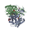
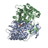
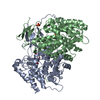
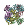
| Title | STRUCTURAL DETERMINATION OF THE A50T:S279G:S280K:V281K:K282E:H283N VARIANT OF CITRATE SYNTHASE FROM E. COLI complexed WITH S-CARBOXYMETHYL-COA | |||||||||
 Components Components | Citrate synthase | |||||||||
 Keywords Keywords | TRANSFERASE / CITRATE SYNTHASE / GRAM-NEGATIVE BACTERIA / ALLOSTERY / OXALOACETATE / ACETYL-COA / NADH / PROTEIN FOLDING / S-CARBOXYMETHYL-COA / ALLOSTERIC ENZYME / TRICARBOXYLIC ACID CYCLE | |||||||||
| Function / homology |  Function and homology information Function and homology informationcitrate synthase (unknown stereospecificity) / citrate synthase activity / NADH binding / protein hexamerization / tricarboxylic acid cycle / identical protein binding / cytosol Similarity search - Function | |||||||||
| Biological species |  | |||||||||
| Method |  X-RAY DIFFRACTION / SYNCHROTRON / MOLECULAR REPLACEMENT / Resolution: 2.7 Å X-RAY DIFFRACTION / SYNCHROTRON / MOLECULAR REPLACEMENT / Resolution: 2.7 Å | |||||||||
 Authors Authors | Maurus, R. / Brayer, G.D. | |||||||||
 Citation Citation | Journal: Biochim.Biophys.Acta / Year: 2013 Title: Enzyme-substrate complexes of allosteric citrate synthase: Evidence for a novel intermediate in substrate binding. Authors: Duckworth, H.W. / Nguyen, N.T. / Gao, Y. / Donald, L.J. / Maurus, R. / Ayed, A. / Bruneau, B. / Brayer, G.D. | |||||||||
| History |
|
- Structure visualization
| Structure viewer | Molecule: MolmilJmol/JSmol |
|---|
- Downloads & links
-Download
| PDBx/mmCIF format | 4jae.cif.gz | 183.1 KB | Display | PDBx/mmCIF format |
|---|---|---|---|---|
| PDB format | pdb4jae.ent.gz | 145.8 KB | Display | PDB format |
| PDBx/mmJSON format | 4jae.json.gz | Tree view | PDBx/mmJSON format | |
| Others |  Other downloads Other downloads |
-Validation report
| Arichive directory | https://data.pdbj.org/pub/pdb/validation_reports/ja/4jaeftp://data.pdbj.org/pub/pdb/validation_reports/ja/4jae | HTTPS FTP |
|---|
-Related structure data
| Related structure data |  4jadC 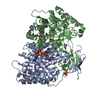 4jafC  4jagC  1k3p C: citing same article ( S: Starting model for refinement |
|---|---|
| Similar structure data |


-Links
 PDBj
PDBj

- Assembly
| Deposited unit | 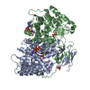
| ||||||||
|---|---|---|---|---|---|---|---|---|---|
| 1 | 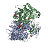
| ||||||||
| Unit cell |
|
-Components
| #1: Protein | Mass: 47994.785 Da / Num. of mol.: 2 / Mutation: A50T, S279G, S280K, V281K, K282E, H283N Source method: isolated from a genetically manipulated source Source: (gene. exp.) #2: Chemical | ChemComp-SO4 /   Mass: 96.063 Da / Num. of mol.: 4 / Source method: obtained synthetically / Formula: SO4 Mass: 96.063 Da / Num. of mol.: 4 / Source method: obtained synthetically / Formula: SO4#3: Chemical |   Mass: 825.570 Da / Num. of mol.: 2 / Source method: obtained synthetically / Formula: C23H38N7O18P3S Mass: 825.570 Da / Num. of mol.: 2 / Source method: obtained synthetically / Formula: C23H38N7O18P3S#4: Water | ChemComp-HOH / |  Mass: 18.015 Da / Num. of mol.: 227 / Source method: isolated from a natural source / Formula: H2O Mass: 18.015 Da / Num. of mol.: 227 / Source method: isolated from a natural source / Formula: H2OSequence details | ASPARTATE AT POSITION 10 CHAIN A AND 1010 CHAIN B IS A POST-TRANSLATIONAL MODIFICATION OF ASN THAT ...ASPARTATE AT POSITION 10 CHAIN A AND 1010 CHAIN B IS A POST-TRANSLATIO | |
|---|
-Experimental details
-Experiment
| Experiment | Method: X-RAY DIFFRACTION / Number of used crystals: 1 |
|---|
- Sample preparation
| Crystal | Density Matthews: 4.27 Å3/Da / Density % sol: 71.2 % |
|---|---|
| Crystal grow | Temperature: 298 K / Method: vapor diffusion, hanging drop / pH: 7.5 Details: 2% (V/V) PEG400, 2.0-2.3 M AMMONIUM SULFATE, 0.1 M NA HEPES, PH 7.5, VAPOR DIFFUSION, HANGING DROP, TEMPERATURE 298K |
-Data collection
| Diffraction | Mean temperature: 100 K |
|---|---|
| Diffraction source | Source: SYNCHROTRON / Site: SSRL  / Beamline: BL7-1 / Wavelength: 0.97 Å / Beamline: BL7-1 / Wavelength: 0.97 Å |
| Detector | Type: ADSC QUANTUM 315r / Detector: CCD / Date: Feb 28, 2008 / Details: VERTICAL FOCUSING MIRROR |
| Radiation | Monochromator: SIDE SCATTERING I-BEAM BENT SINGLE CRYSTAL / Protocol: SINGLE WAVELENGTH / Monochromatic (M) / Laue (L): M / Scattering type: x-ray |
| Radiation wavelength | Wavelength: 0.97 Å / Relative weight: 1 |
| Reflection | Resolution: 2.7→30 Å / Num. obs: 42859 / % possible obs: 98.1 % / Redundancy: 2.8 % / Rmerge(I) obs: 0.086 / Net I/σ(I): 7 |
- Processing
| Software |
| ||||||||||||||||||||||||||||||||||||||||||||||||||||||||||||
|---|---|---|---|---|---|---|---|---|---|---|---|---|---|---|---|---|---|---|---|---|---|---|---|---|---|---|---|---|---|---|---|---|---|---|---|---|---|---|---|---|---|---|---|---|---|---|---|---|---|---|---|---|---|---|---|---|---|---|---|---|---|
| Refinement | Method to determine structure: MOLECULAR REPLACEMENT Starting model: 1K3P 1k3p Resolution: 2.7→30 Å / σ(F): 0 / Stereochemistry target values: ENGH & HUBER
| ||||||||||||||||||||||||||||||||||||||||||||||||||||||||||||
| Solvent computation | Bsol: 76.42 Å2 | ||||||||||||||||||||||||||||||||||||||||||||||||||||||||||||
| Displacement parameters | Biso mean: 48.57 Å2 | ||||||||||||||||||||||||||||||||||||||||||||||||||||||||||||
| Refinement step | Cycle: LAST / Resolution: 2.7→30 Å
| ||||||||||||||||||||||||||||||||||||||||||||||||||||||||||||
| Refine LS restraints |
| ||||||||||||||||||||||||||||||||||||||||||||||||||||||||||||
| Xplor file |
|
