 Movie
Movie Controller

[English] 日本語
 Yorodumi
- PDB-1owc: Three Dimensional Structure Analysis Of The R109L Variant of the ... -
+ Open data

- Basic information
| Entry | Database: PDB / ID: 1owc | ||||||
|---|---|---|---|---|---|---|---|
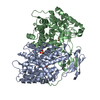
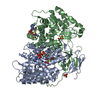
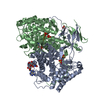
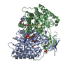
| Title | Three Dimensional Structure Analysis Of The R109L Variant of the Type II Citrate Synthase From E. Coli | ||||||
 Components Components | Citrate synthase | ||||||
 Keywords Keywords | TRANSFERASE / Allostery / NADH / Type II Citrate Synthase / E. Coli / R109L | ||||||
| Function / homology |  Function and homology information Function and homology informationcitrate synthase (unknown stereospecificity) / citrate synthase activity / NADH binding / protein hexamerization / tricarboxylic acid cycle / identical protein binding / cytosol Similarity search - Function | ||||||
| Biological species |  | ||||||
| Method |  X-RAY DIFFRACTION / MOLECULAR REPLACEMENT / Resolution: 2.2 Å X-RAY DIFFRACTION / MOLECULAR REPLACEMENT / Resolution: 2.2 Å | ||||||
 Authors Authors | Stokell, D.J. / Donald, L.J. / Maurus, R. / Nguyen, N.T. / Sadler, G. / Choudhary, K. / Hultin, P.G. / Brayer, G.D. / Duckworth, H.W. | ||||||
 Citation Citation | Journal: J.Biol.Chem. / Year: 2003 Title: Probing the roles of key residues in the unique regulatory NADH binding site of type II citrate synthase of Escherichia coli. Authors: Stokell, D.J. / Donald, L.J. / Maurus, R. / Nguyen, N.T. / Sadler, G. / Choudhary, K. / Hultin, P.G. / Brayer, G.D. / Duckworth, H.W. | ||||||
| History |
|
- Structure visualization
| Structure viewer | Molecule: MolmilJmol/JSmol |
|---|
- Downloads & links
-Download
| PDBx/mmCIF format | 1owc.cif.gz | 185.9 KB | Display | PDBx/mmCIF format |
|---|---|---|---|---|
| PDB format | pdb1owc.ent.gz | 147.2 KB | Display | PDB format |
| PDBx/mmJSON format | 1owc.json.gz | Tree view | PDBx/mmJSON format | |
| Others |  Other downloads Other downloads |
-Validation report
| Arichive directory | https://data.pdbj.org/pub/pdb/validation_reports/ow/1owcftp://data.pdbj.org/pub/pdb/validation_reports/ow/1owc | HTTPS FTP |
|---|
-Related structure data
| Related structure data |  1owbC  1k3p C: citing same article ( S: Starting model for refinement |
|---|---|
| Similar structure data |


-Links
 PDBj
PDBj

- Assembly
| Deposited unit | 
| ||||||||
|---|---|---|---|---|---|---|---|---|---|
| 1 | 
| ||||||||
| Unit cell |
|
-Components
| #1: Protein | Mass: 48080.957 Da / Num. of mol.: 2 / Mutation: R109L Source method: isolated from a genetically manipulated source Source: (gene. exp.) #2: Chemical | ChemComp-SO4 /   Mass: 96.063 Da / Num. of mol.: 6 / Source method: obtained synthetically / Formula: SO4 Mass: 96.063 Da / Num. of mol.: 6 / Source method: obtained synthetically / Formula: SO4#3: Water | ChemComp-HOH / |  Mass: 18.015 Da / Num. of mol.: 511 / Source method: isolated from a natural source / Formula: H2O Mass: 18.015 Da / Num. of mol.: 511 / Source method: isolated from a natural source / Formula: H2O |
|---|
-Experimental details
-Experiment
| Experiment | Method: X-RAY DIFFRACTION |
|---|
- Sample preparation
| Crystal | Density Matthews: 4.23 Å3/Da / Density % sol: 70.94 % |
|---|---|
| Crystal grow | Method: vapor diffusion, hanging drop / Details: VAPOR DIFFUSION, HANGING DROP |
-Data collection
| Diffraction | Mean temperature: 100 K |
|---|---|
| Diffraction source | Source: ROTATING ANODE / Type: RIGAKU RU300 / Wavelength: 1.5418 Å |
| Detector | Type: RIGAKU RAXIS IIC / Detector: IMAGE PLATE / Date: Jul 3, 2000 / Details: osmic mirror |
| Radiation | Monochromator: Osmic Mirrors / Protocol: SINGLE WAVELENGTH / Monochromatic (M) / Laue (L): M / Scattering type: x-ray |
| Radiation wavelength | Wavelength: 1.5418 Å / Relative weight: 1 |
| Reflection | Highest resolution: 2.2 Å / Num. obs: 72746 / % possible obs: 88 % / Observed criterion σ(F): 0 / Observed criterion σ(I): 0 / Redundancy: 4.6 % / Rmerge(I) obs: 0.094 |
- Processing
| Software |
| ||||||||||||
|---|---|---|---|---|---|---|---|---|---|---|---|---|---|
| Refinement | Method to determine structure: MOLECULAR REPLACEMENT Starting model: PDB ENTRY 1K3P 1k3p Resolution: 2.2→10 Å / Stereochemistry target values: Engh & Huber /
| ||||||||||||
| Refinement step | Cycle: LAST / Resolution: 2.2→10 Å
| ||||||||||||
| Refine LS restraints |
|
