 Movie
Movie Controller

[English] 日本語
 Yorodumi
- PDB-2dgm: Crystal structure of Escherichia coli GadB in complex with iodide -
+ Open data

- Basic information
| Entry | Database: PDB / ID: 2dgm | ||||||
|---|---|---|---|---|---|---|---|
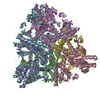
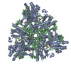
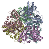
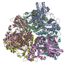
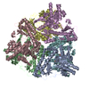
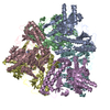
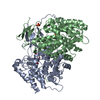
| Title | Crystal structure of Escherichia coli GadB in complex with iodide | ||||||
 Components Components | Glutamate decarboxylase beta | ||||||
 Keywords Keywords | LYASE / GadB complexed with iodide | ||||||
| Function / homology |  Function and homology information Function and homology informationglutamate decarboxylase / glutamate decarboxylase activity / intracellular pH elevation / L-glutamate catabolic process / pyridoxal phosphate binding / membrane / cytosol Similarity search - Function | ||||||
| Biological species |  | ||||||
| Method |  X-RAY DIFFRACTION / SYNCHROTRON / MOLECULAR REPLACEMENT / Resolution: 1.95 Å X-RAY DIFFRACTION / SYNCHROTRON / MOLECULAR REPLACEMENT / Resolution: 1.95 Å | ||||||
 Authors Authors | Gruetter, M.G. / Capitani, G. / Gut, H. | ||||||
 Citation Citation | Journal: Embo J. / Year: 2006 Title: Escherichia coli acid resistance: pH-sensing, activation by chloride and autoinhibition in GadB Authors: Gut, H. / Pennacchietti, E. / John, R.A. / Bossa, F. / Capitani, G. / De Biase, D. / Gruetter, M.G. #1: Journal: Embo J. / Year: 2003Title: Crystal structure and functional analysis of Escherichia coli glutamate decarboxylase Authors: Capitani, G. / De Biase, D. / Aurizi, C. / Gut, H. / Bossa, F. / Gruetter, M.G. | ||||||
| History |
|
- Structure visualization
| Structure viewer | Molecule: MolmilJmol/JSmol |
|---|
- Downloads & links
-Download
| PDBx/mmCIF format | 2dgm.cif.gz | 570 KB | Display | PDBx/mmCIF format |
|---|---|---|---|---|
| PDB format | pdb2dgm.ent.gz | 464.8 KB | Display | PDB format |
| PDBx/mmJSON format | 2dgm.json.gz | Tree view | PDBx/mmJSON format | |
| Others |  Other downloads Other downloads |
-Validation report
| Arichive directory | https://data.pdbj.org/pub/pdb/validation_reports/dg/2dgmftp://data.pdbj.org/pub/pdb/validation_reports/dg/2dgm | HTTPS FTP |
|---|
-Related structure data
| Related structure data | 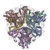 2dgkC  2dglC  1pmmS S: Starting model for refinement C: citing same article ( |
|---|---|
| Similar structure data |


-Links
 PDBj
PDBj

- Assembly
| Deposited unit | 
| ||||||||
|---|---|---|---|---|---|---|---|---|---|
| 1 |
| ||||||||
| Unit cell |
|
-Components
-Protein , 1 types, 6 molecules ABCDEF
| #1: Protein | Mass: 52727.957 Da / Num. of mol.: 6 Source method: isolated from a genetically manipulated source Source: (gene. exp.) |
|---|
-Non-polymers , 6 types, 1976 molecules 


| #2: Chemical | ChemComp-IOD /  Mass: 126.904 Da / Num. of mol.: 32 / Source method: obtained synthetically / Formula: I Mass: 126.904 Da / Num. of mol.: 32 / Source method: obtained synthetically / Formula: I#3: Chemical | ChemComp-PLP /  Mass: 247.142 Da / Num. of mol.: 6 / Source method: obtained synthetically / Formula: C8H10NO6P Mass: 247.142 Da / Num. of mol.: 6 / Source method: obtained synthetically / Formula: C8H10NO6P#4: Chemical | ChemComp-FMT /  Mass: 46.025 Da / Num. of mol.: 13 / Source method: obtained synthetically / Formula: CH2O2 Mass: 46.025 Da / Num. of mol.: 13 / Source method: obtained synthetically / Formula: CH2O2#5: Chemical | ChemComp-ACY / |  Mass: 60.052 Da / Num. of mol.: 1 / Source method: obtained synthetically / Formula: C2H4O2 Mass: 60.052 Da / Num. of mol.: 1 / Source method: obtained synthetically / Formula: C2H4O2#6: Chemical | ChemComp-PEG / |  Mass: 106.120 Da / Num. of mol.: 1 / Source method: obtained synthetically / Formula: C4H10O3 Mass: 106.120 Da / Num. of mol.: 1 / Source method: obtained synthetically / Formula: C4H10O3#7: Water | ChemComp-HOH / | Mass: 18.015 Da / Num. of mol.: 1923 / Source method: isolated from a natural source / Formula: H2O |
|---|
-Experimental details
-Experiment
| Experiment | Method: X-RAY DIFFRACTION / Number of used crystals: 1 |
|---|
- Sample preparation
| Crystal | Density Matthews: 2.33 Å3/Da / Density % sol: 47.13 % |
|---|---|
| Crystal grow | Temperature: 293 K / Method: vapor diffusion, sitting drop / pH: 4.6 Details: 0.135M sodium acetate, 0.7M sodium formate, 13% PEG 4000, pH 4.6, VAPOR DIFFUSION, SITTING DROP, temperature 293K |
-Data collection
| Diffraction | Mean temperature: 90 K |
|---|---|
| Diffraction source | Source: SYNCHROTRON / Site: SLS  / Beamline: X06SA / Wavelength: 1.3 Å / Beamline: X06SA / Wavelength: 1.3 Å |
| Detector | Type: MARRESEARCH / Detector: CCD / Date: May 2, 2003 / Details: Dynamically bendable mirror |
| Radiation | Monochromator: LN2 cooled fixed-exit Si(111) monochromator / Protocol: SINGLE WAVELENGTH / Monochromatic (M) / Laue (L): M / Scattering type: x-ray |
| Radiation wavelength | Wavelength: 1.3 Å / Relative weight: 1 |
| Reflection | Resolution: 1.95→40 Å / Num. obs: 191280 / % possible obs: 92.7 % / Observed criterion σ(I): -3 / Redundancy: 1.8 % / Biso Wilson estimate: 20.9 Å2 / Rsym value: 0.12 / Net I/σ(I): 5.9 |
| Reflection shell | Resolution: 1.95→2.02 Å / Redundancy: 1.5 % / Mean I/σ(I) obs: 2.7 / Num. unique all: 17342 / Rsym value: 0.26 / % possible all: 84.4 |
- Processing
| Software |
| ||||||||||||||||||||
|---|---|---|---|---|---|---|---|---|---|---|---|---|---|---|---|---|---|---|---|---|---|
| Refinement | Method to determine structure: MOLECULAR REPLACEMENT Starting model: PDB entry 1PMM Resolution: 1.95→40 Å / Isotropic thermal model: Isotropic / Cross valid method: THROUGHOUT / σ(F): 0 / Stereochemistry target values: Engh & Huber
| ||||||||||||||||||||
| Displacement parameters | Biso mean: 21 Å2 | ||||||||||||||||||||
| Refine analyze |
| ||||||||||||||||||||
| Refinement step | Cycle: LAST / Resolution: 1.95→40 Å
| ||||||||||||||||||||
| Refine LS restraints |
| ||||||||||||||||||||
| LS refinement shell | Resolution: 1.95→2.02 Å
|
