 Movie
Movie Controller

[English] 日本語
 Yorodumi
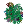
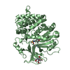
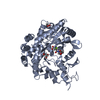
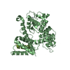
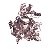
- PDB-1r2x: Coordinates of L11 with 58nts of 23S rRNA fitted into the cryo-EM... -
+ Open data

- Basic information
| Entry | Database: PDB / ID: 1r2x | ||||||
|---|---|---|---|---|---|---|---|
| Title | Coordinates of L11 with 58nts of 23S rRNA fitted into the cryo-EM map of EF-Tu ternary complex (GDP.Kirromycin) bound 70S ribosome | ||||||
 Components Components |
| ||||||
 Keywords Keywords | RNA BINDING PROTEIN/RNA / rna / ribosomal protein / RNA BINDING PROTEIN-RNA COMPLEX | ||||||
| Function / homology |  Function and homology information Function and homology informationlarge ribosomal subunit rRNA binding / cytosolic large ribosomal subunit / structural constituent of ribosome / translation Similarity search - Function | ||||||
| Biological species |   Thermotoga maritima (bacteria) Thermotoga maritima (bacteria) | ||||||
| Method | ELECTRON MICROSCOPY / single particle reconstruction / cryo EM / Resolution: 9 Å | ||||||
 Authors Authors | Valle, M. / Zavialov, A. / Li, W. / Stagg, S.M. / Sengupta, J. / Nielsen, R.C. / Nissen, P. / Harvey, S.C. / Ehrenberg, M. / Frank, J. | ||||||
 Citation Citation | Journal: Nat Struct Biol / Year: 2003 Title: Incorporation of aminoacyl-tRNA into the ribosome as seen by cryo-electron microscopy. Authors: Mikel Valle / Andrey Zavialov / Wen Li / Scott M Stagg / Jayati Sengupta / Rikke C Nielsen / Poul Nissen / Stephen C Harvey / Måns Ehrenberg / Joachim Frank /  Abstract: Aminoacyl-tRNAs (aa-tRNAs) are delivered to the ribosome as part of the ternary complex of aa-tRNA, elongation factor Tu (EF-Tu) and GTP. Here, we present a cryo-electron microscopy (cryo-EM) study, ...Aminoacyl-tRNAs (aa-tRNAs) are delivered to the ribosome as part of the ternary complex of aa-tRNA, elongation factor Tu (EF-Tu) and GTP. Here, we present a cryo-electron microscopy (cryo-EM) study, at a resolution of approximately 9 A, showing that during the incorporation of the aa-tRNA into the 70S ribosome of Escherichia coli, the flexibility of aa-tRNA allows the initial codon recognition and its accommodation into the ribosomal A site. In addition, a conformational change observed in the GTPase-associated center (GAC) of the ribosomal 50S subunit may provide the mechanism by which the ribosome promotes a relative movement of the aa-tRNA with respect to EF-Tu. This relative rearrangement seems to facilitate codon recognition by the incoming aa-tRNA, and to provide the codon-anticodon recognition-dependent signal for the GTPase activity of EF-Tu. From these new findings we propose a mechanism that can explain the sequence of events during the decoding of mRNA on the ribosome. | ||||||
| History |
| ||||||
| Remark 999 | SEQUENCE THE PROTEIN STRUCTURE CONTAINS CA ATOMS and THE RNA STRUCTURE CONTAINS P ATOMS ONLY |
- Structure visualization
| Movie |
Movie viewer |
|---|---|
| Structure viewer | Molecule: MolmilJmol/JSmol |
- Downloads & links
-Download
| PDBx/mmCIF format | 1r2x.cif.gz | 19.2 KB | Display | PDBx/mmCIF format |
|---|---|---|---|---|
| PDB format | pdb1r2x.ent.gz | 7.4 KB | Display | PDB format |
| PDBx/mmJSON format | 1r2x.json.gz | Tree view | PDBx/mmJSON format | |
| Others |  Other downloads Other downloads |
-Validation report
| Arichive directory | https://data.pdbj.org/pub/pdb/validation_reports/r2/1r2xftp://data.pdbj.org/pub/pdb/validation_reports/r2/1r2x | HTTPS FTP |
|---|
-Related structure data
| Related structure data |  1055MC  1056C  1395C 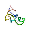 1qzaC 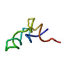 1qzbC 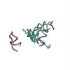 1qzcC 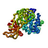 1qzdC 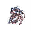 1r2wC C: citing same article ( M: map data used to model this data |
|---|---|
| Similar structure data |
-Links
 PDBj
PDBj


- Assembly
| Deposited unit | 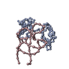
|
|---|---|
| 1 |
|
-Components
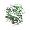
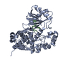
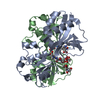
| #1: RNA chain | Mass: 18693.145 Da / Num. of mol.: 1 / Source method: obtained synthetically |
|---|---|
| #2: Protein | Mass: 15109.841 Da / Num. of mol.: 1 / Source method: isolated from a natural source / Source: (natural) Thermotoga maritima (bacteria) / References: UniProt: P29395 |
-Experimental details
-Experiment
| Experiment | Method: ELECTRON MICROSCOPY |
|---|---|
| EM experiment | Aggregation state: PARTICLE / 3D reconstruction method: single particle reconstruction |
- Sample preparation
| Component |
| ||||||||||||||||||||
|---|---|---|---|---|---|---|---|---|---|---|---|---|---|---|---|---|---|---|---|---|---|
| Buffer solution | Name: Polymix buffer / pH: 7.5 / Details: Polymix buffer | ||||||||||||||||||||
| Specimen | Embedding applied: NO / Shadowing applied: NO / Staining applied: NO / Vitrification applied: YES | ||||||||||||||||||||
| Specimen support | Details: Quantifoil holley carbon coated grids | ||||||||||||||||||||
| Vitrification | Cryogen name: ETHANE / Details: Rapid freezing in liquid ethane | ||||||||||||||||||||
| Crystal grow | *PLUS Method: electron microscopy / Details: electron microscopy |
- Electron microscopy imaging
| Experimental equipment |  Model: Tecnai F20 / Image courtesy: FEI Company |
|---|---|
| Microscopy | Model: FEI TECNAI F20 |
| Electron gun | Electron source:  FIELD EMISSION GUN / Accelerating voltage: 200 kV / Illumination mode: FLOOD BEAM FIELD EMISSION GUN / Accelerating voltage: 200 kV / Illumination mode: FLOOD BEAM |
| Electron lens | Mode: BRIGHT FIELD / Nominal magnification: 50000 X / Nominal defocus max: 4000 nm / Nominal defocus min: 2000 nm |
| Specimen holder | Temperature: 93 K / Tilt angle max: 0 ° / Tilt angle min: 0 ° |
| Image recording | Electron dose: 200 e/Å2 / Film or detector model: KODAK SO-163 FILM |
- Processing
| Symmetry | Point symmetry: C1 (asymmetric) | ||||||||||||
|---|---|---|---|---|---|---|---|---|---|---|---|---|---|
| 3D reconstruction | Method: Reference based alignment / Resolution: 9 Å / Num. of particles: 75996 / Actual pixel size: 2.82 Å / Magnification calibration: TMV / Symmetry type: POINT | ||||||||||||
| Atomic model building | Protocol: OTHER / Details: METHOD--manual using O | ||||||||||||
| Refinement step | Cycle: LAST
|
