Movie
Movie Controller


+ Open data

- Basic information
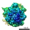
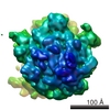
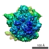
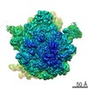
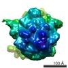
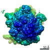
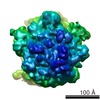
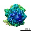
| Entry | Database: EMDB / ID: EMD-1055 | |||||||||
|---|---|---|---|---|---|---|---|---|---|---|
| Title | Locking and unlocking of ribosomal motions. | |||||||||
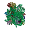
 Map data Map data | 70S + fMet-tRNA + PhetRNA + EF-Tu + GDP + kir mRNA codes for MP-stop | |||||||||
 Sample Sample |
| |||||||||
| Function / homology |  Function and homology information Function and homology informationguanyl-nucleotide exchange factor complex / protein-synthesizing GTPase / guanosine tetraphosphate binding / stringent response / negative regulation of cytoplasmic translational initiation / transcription antitermination factor activity, RNA binding / ornithine decarboxylase inhibitor activity / translational elongation / misfolded RNA binding / Group I intron splicing ...guanyl-nucleotide exchange factor complex / protein-synthesizing GTPase / guanosine tetraphosphate binding / stringent response / negative regulation of cytoplasmic translational initiation / transcription antitermination factor activity, RNA binding / ornithine decarboxylase inhibitor activity / translational elongation / misfolded RNA binding / Group I intron splicing / RNA folding / translation elongation factor activity / translational termination / transcriptional attenuation / endoribonuclease inhibitor activity / positive regulation of ribosome biogenesis / RNA-binding transcription regulator activity / four-way junction DNA binding / negative regulation of cytoplasmic translation / DnaA-L2 complex / translation repressor activity / negative regulation of translational initiation / negative regulation of DNA-templated DNA replication initiation / mRNA regulatory element binding translation repressor activity / positive regulation of RNA splicing / regulation of DNA-templated transcription elongation / transcription elongation factor complex / response to reactive oxygen species / cytosolic ribosome assembly / ribosome assembly / assembly of large subunit precursor of preribosome / transcription antitermination / DNA endonuclease activity / regulation of cell growth / translational initiation / DNA-templated transcription termination / response to radiation / maintenance of translational fidelity / mRNA 5'-UTR binding / regulation of translation / large ribosomal subunit / transferase activity / ribosomal small subunit assembly / ribosome binding / ribosome biogenesis / ribosomal small subunit biogenesis / 5S rRNA binding / ribosomal large subunit assembly / small ribosomal subunit / small ribosomal subunit rRNA binding / large ribosomal subunit rRNA binding / cytosolic small ribosomal subunit / cytosolic large ribosomal subunit / cytoplasmic translation / tRNA binding / negative regulation of translation / rRNA binding / structural constituent of ribosome / ribosome / translation / ribonucleoprotein complex / response to antibiotic / negative regulation of DNA-templated transcription / hydrolase activity / mRNA binding / GTPase activity / GTP binding / magnesium ion binding / DNA binding / RNA binding / zinc ion binding / membrane / plasma membrane / cytosol / cytoplasm Similarity search - Function | |||||||||
| Biological species |  | |||||||||
| Method | single particle reconstruction / cryo EM / negative staining / Resolution: 9.0 Å | |||||||||
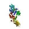
 Authors Authors | Valle M / Zavialov A / Li W / Stagg SM / Sengupta J / Nielsen RC / Nissen P / Hervey SC / Ehrenberg M / Frank J | |||||||||
 Citation Citation | Journal: Nat Struct Biol / Year: 2003 Title: Incorporation of aminoacyl-tRNA into the ribosome as seen by cryo-electron microscopy. Authors: Mikel Valle / Andrey Zavialov / Wen Li / Scott M Stagg / Jayati Sengupta / Rikke C Nielsen / Poul Nissen / Stephen C Harvey / Måns Ehrenberg / Joachim Frank /  Abstract: Aminoacyl-tRNAs (aa-tRNAs) are delivered to the ribosome as part of the ternary complex of aa-tRNA, elongation factor Tu (EF-Tu) and GTP. Here, we present a cryo-electron microscopy (cryo-EM) study, ...Aminoacyl-tRNAs (aa-tRNAs) are delivered to the ribosome as part of the ternary complex of aa-tRNA, elongation factor Tu (EF-Tu) and GTP. Here, we present a cryo-electron microscopy (cryo-EM) study, at a resolution of approximately 9 A, showing that during the incorporation of the aa-tRNA into the 70S ribosome of Escherichia coli, the flexibility of aa-tRNA allows the initial codon recognition and its accommodation into the ribosomal A site. In addition, a conformational change observed in the GTPase-associated center (GAC) of the ribosomal 50S subunit may provide the mechanism by which the ribosome promotes a relative movement of the aa-tRNA with respect to EF-Tu. This relative rearrangement seems to facilitate codon recognition by the incoming aa-tRNA, and to provide the codon-anticodon recognition-dependent signal for the GTPase activity of EF-Tu. From these new findings we propose a mechanism that can explain the sequence of events during the decoding of mRNA on the ribosome. | |||||||||
| History |
|
- Structure visualization
| Movie |
Movie viewer |
|---|---|
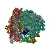
| Structure viewer | EM map: SurfViewMolmilJmol/JSmol |
| Supplemental images |

- Downloads & links
-EMDB archive
| Map data | emd_1055.map.gz | 7.8 MB | EMDB map data format | |
|---|---|---|---|---|
| Header (meta data) | emd-1055-v30.xmlemd-1055.xml | 12.7 KB 12.7 KB | Display Display | EMDB header |
| Images | 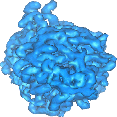 1055.gif 1055.gif | 80.1 KB | ||
| Archive directory |  http://ftp.pdbj.org/pub/emdb/structures/EMD-1055ftp://ftp.pdbj.org/pub/emdb/structures/EMD-1055 http://ftp.pdbj.org/pub/emdb/structures/EMD-1055ftp://ftp.pdbj.org/pub/emdb/structures/EMD-1055 | HTTPS FTP |
-Related structure data
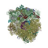
| Related structure data | 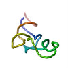 1qzaMC  1qzdMC  1r2xMC 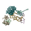 3ep2M  4v65M 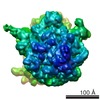 1056C 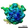 1395C 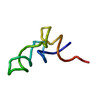 1qzbC 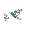 1qzcC  1r2wC M: atomic model generated by this map C: citing same article ( |
|---|---|
| Similar structure data |

-Links
| EMDB pages | EMDB (EBI/PDBe) / EMDataResource |
|---|---|
| Related items in Molecule of the Month |


-Map
| File | Download / File: emd_1055.map.gz / Format: CCP4 / Size: 8.2 MB / Type: IMAGE STORED AS FLOATING POINT NUMBER (4 BYTES) | ||||||||||||||||||||||||||||||||||||||||||||||||||||||||||||||||||||
|---|---|---|---|---|---|---|---|---|---|---|---|---|---|---|---|---|---|---|---|---|---|---|---|---|---|---|---|---|---|---|---|---|---|---|---|---|---|---|---|---|---|---|---|---|---|---|---|---|---|---|---|---|---|---|---|---|---|---|---|---|---|---|---|---|---|---|---|---|---|
| Annotation | 70S + fMet-tRNA + PhetRNA + EF-Tu + GDP + kir mRNA codes for MP-stop | ||||||||||||||||||||||||||||||||||||||||||||||||||||||||||||||||||||
| Projections & slices | Image control
Images are generated by Spider. | ||||||||||||||||||||||||||||||||||||||||||||||||||||||||||||||||||||
| Voxel size | X=Y=Z: 2.82 Å | ||||||||||||||||||||||||||||||||||||||||||||||||||||||||||||||||||||
| Density |
| ||||||||||||||||||||||||||||||||||||||||||||||||||||||||||||||||||||
| Symmetry | Space group: 1 | ||||||||||||||||||||||||||||||||||||||||||||||||||||||||||||||||||||
| Details | EMDB XML:
CCP4 map header:
| ||||||||||||||||||||||||||||||||||||||||||||||||||||||||||||||||||||
 Z (Sec.)
 Y (Row.)
 X (Col.)


-Supplemental data
- Sample components
-Entire : 70S ribosome from E. coli complex 70S-fMet-tRNA-Phe-tRNA-EF-Tu-GD...
| Entire | Name: 70S ribosome from E. coli complex 70S-fMet-tRNA-Phe-tRNA-EF-Tu-GDP-kirromycin. |
|---|---|
| Components |
|
-Supramolecule #1000: 70S ribosome from E. coli complex 70S-fMet-tRNA-Phe-tRNA-EF-Tu-GD...
| Supramolecule | Name: 70S ribosome from E. coli complex 70S-fMet-tRNA-Phe-tRNA-EF-Tu-GDP-kirromycin. type: sample / ID: 1000 / Number unique components: 5 |
|---|
-Supramolecule #1: 70S from E. coli
| Supramolecule | Name: 70S from E. coli / type: complex / ID: 1 / Name.synonym: ribosome / Recombinant expression: No / Ribosome-details: ribosome-prokaryote: ALL |
|---|---|
| Source (natural) | Organism: |
-Macromolecule #1: fMet-tRNAfMet
| Macromolecule | Name: fMet-tRNAfMet / type: rna / ID: 1 / Name.synonym: trna / Classification: OTHER / Structure: DOUBLE HELIX / Synthetic?: No |
|---|---|
| Source (natural) | Organism: |
-Macromolecule #2: Phe-tRNAPhe
| Macromolecule | Name: Phe-tRNAPhe / type: rna / ID: 2 / Name.synonym: trna / Classification: OTHER / Structure: DOUBLE HELIX / Synthetic?: No |
|---|---|
| Source (natural) | Organism: |
-Macromolecule #4: deacylated tRNA
| Macromolecule | Name: deacylated tRNA / type: rna / ID: 4 / Name.synonym: trna / Classification: OTHER / Structure: DOUBLE HELIX / Synthetic?: No |
|---|---|
| Source (natural) | Organism: |
-Macromolecule #3: Elongation Factor Tu
| Macromolecule | Name: Elongation Factor Tu / type: protein_or_peptide / ID: 3 / Name.synonym: Elongation Factor / Number of copies: 1 / Recombinant expression: No |
|---|---|
| Source (natural) | Organism: |
-Experimental details
-Structure determination
| Method | negative staining, cryo EM |
|---|---|
 Processing Processing | single particle reconstruction |
| Aggregation state | particle |
-Sample preparation
| Buffer | pH: 7.5 Details: 5 mM potassium phosphate, 5 mM magnesium acetate, 5 mM ammonium chloride, 95 mM potassium chloride, 0.5 mM calcium chloride, 8 mM putrescine, 1 mM spermidine, and 1 mM dithioerythritol |
|---|---|
| Staining | Type: NEGATIVE / Details: Cryo-electron microscopy. No stain. |
| Grid | Details: 300 mesh Quantifoil R2/4 |
| Vitrification | Cryogen name: ETHANE / Chamber temperature: 90 K / Method: two-face blotting for 1 second |
- Electron microscopy
| Microscope | FEI TECNAI F20 |
|---|---|
| Temperature | Average: 90 K |
| Alignment procedure | Legacy - Astigmatism: corrected at 175,000 |
| Image recording | Category: FILM / Film or detector model: KODAK SO-163 FILM / Digitization - Scanner: ZEISS SCAI / Digitization - Sampling interval: 14 µm / Number real images: 50 / Average electron dose: 15 e/Å2 / Od range: 1 / Bits/pixel: 16 |
| Tilt angle min | 0 |
| Tilt angle max | 0 |
| Electron beam | Acceleration voltage: 200 kV / Electron source:  FIELD EMISSION GUN FIELD EMISSION GUN |
| Electron optics | Calibrated magnification: 49650 / Illumination mode: FLOOD BEAM / Imaging mode: BRIGHT FIELD / Cs: 2 mm / Nominal defocus max: 4.0 µm / Nominal defocus min: 2.0 µm / Nominal magnification: 50000 |
| Sample stage | Specimen holder: single tilt cryoholder / Specimen holder model: GATAN LIQUID NITROGEN |
| Experimental equipment |  Model: Tecnai F20 / Image courtesy: FEI Company |
-Image processing
| CTF correction | Details: segregation in defocus groups and correction in volumes |
|---|---|
| Final reconstruction | Applied symmetry - Point group: C1 (asymmetric) / Algorithm: OTHER / Resolution.type: BY AUTHOR / Resolution: 9.0 Å / Resolution method: OTHER / Software - Name: Spider / Number images used: 75996 |
-Atomic model buiding 1
| Initial model | (PDB ID: , ,  1fjf |
|---|---|
| Software | Name: O |
| Details | Protocol: rigid body. Manual fitting in O.The docking of X-ray structures into the cryo-EM maps was made using O and the visualization was performed in IRIS Explorer. EF-G domains were taken from the Thermus thermophilus H573A mutant crystal structure in complex with GDP (PDB code: 1FNM). The coordinates of the ribosomal proteins and rRNAs were taken from published atomic structures (PDB code 1FFK for the 50S subunit; 1FJF for the 30S subunit and the docking was performed using the coordinates as rigid bodies. |
| Refinement | Protocol: RIGID BODY FIT / Target criteria: correlation coeficient |
| Output model | PDB-1qza: PDB-1qzd: PDB-1r2x: PDB-3ep2: PDB-4v65: |