National Institutes of Health/National Institute of General Medical Sciences (NIH/NIGMS)
GM33050
米国
National Institutes of Health/National Heart, Lung, and Blood Institute (NIH/NHLBI)
HL51811
米国
National Institutes of Health/National Institute Of Allergy and Infectious Diseases (NIH/NIAID)
AI1081961
米国
National Institutes of Health/National Institute of General Medical Sciences (NIH/NIGMS)
T32-GM008799
米国
引用
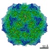
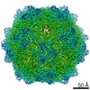
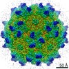
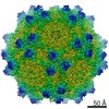
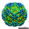
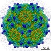
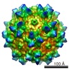
ジャーナル: J Virol / 年: 2016 タイトル: Cryo-electron Microscopy Reconstruction and Stability Studies of the Wild Type and the R432A Variant of Adeno-associated Virus Type 2 Reveal that Capsid Structural Stability Is a Major ...タイトル: Cryo-electron Microscopy Reconstruction and Stability Studies of the Wild Type and the R432A Variant of Adeno-associated Virus Type 2 Reveal that Capsid Structural Stability Is a Major Factor in Genome Packaging. 著者: Lauren M Drouin / Bridget Lins / Maria Janssen / Antonette Bennett / Paul Chipman / Robert McKenna / Weijun Chen / Nicholas Muzyczka / Giovanni Cardone / Timothy S Baker / Mavis Agbandje-McKenna / 要旨: The adeno-associated viruses (AAV) are promising therapeutic gene delivery vectors and better understanding of their capsid assembly and genome packaging mechanism is needed for improved vector ...The adeno-associated viruses (AAV) are promising therapeutic gene delivery vectors and better understanding of their capsid assembly and genome packaging mechanism is needed for improved vector production. Empty AAV capsids assemble in the nucleus prior to genome packaging by virally encoded Rep proteins. To elucidate the capsid determinants of this process, structural differences between wild-type (wt) AAV2 and a packaging deficient variant, AAV2-R432A, were examined using cryo-electron microscopy and three-dimensional image reconstruction both at an ∼5.0-Å resolution (medium) and also at 3.8- and 3.7-Å resolutions (high), respectively. The high resolution structures showed that removal of the arginine side chain in AAV2-R432A eliminated hydrogen bonding interactions, resulting in altered intramolecular and intermolecular interactions propagated from under the 3-fold axis toward the 5-fold channel. Consistent with these observations, differential scanning calorimetry showed an ∼10°C decrease in thermal stability for AAV2-R432A compared to wt-AAV2. In addition, the medium resolution structures revealed differences in the juxtaposition of the less ordered, N-terminal region of their capsid proteins, VP1/2/3. A structural rearrangement in AAV2-R432A repositioned the βA strand region under the icosahedral 2-fold axis rather than antiparallel to the βB strand, eliminating many intramolecular interactions. Thus, a single amino acid substitution can significantly alter the AAV capsid integrity to the extent of reducing its stability and possibly rendering it unable to tolerate the stress of genome packaging. Furthermore, the data show that the 2-, 3-, and 5-fold regions of the capsid contributed to producing the packaging defect and highlight a tight connection between the entire capsid in maintaining packaging efficiency. IMPORTANCE: The mechanism of AAV genome packaging is still poorly understood, particularly with respect to the capsid determinants of the required capsid-Rep interaction. Understanding this mechanism ...IMPORTANCE: The mechanism of AAV genome packaging is still poorly understood, particularly with respect to the capsid determinants of the required capsid-Rep interaction. Understanding this mechanism may aid in the improvement of AAV packaging efficiency, which is currently ∼1:10 (10%) genome packaged to empty capsid in vector preparations. This report identifies regions of the AAV capsid that play roles in genome packaging and that may be important for Rep recognition. It also demonstrates the need to maintain capsid stability for the success of this process. This information is important for efforts to improve AAV genome packaging and will also inform the engineering of AAV capsid variants for improved tropism, specific tissue targeting, and host antibody escape by defining amino acids that cannot be altered without detriment to infectious vector production.
 ムービー
ムービー コントローラー


 データを開く
 基本情報
基本情報 マップデータ
 試料
試料 キーワード
キーワード 機能・相同性情報
 Adeno-associated virus - 2 (アデノ随伴ウイルス)
 データ登録者
 米国, 4件
  引用
引用 構造の表示
構造の表示
 ダウンロードとリンク
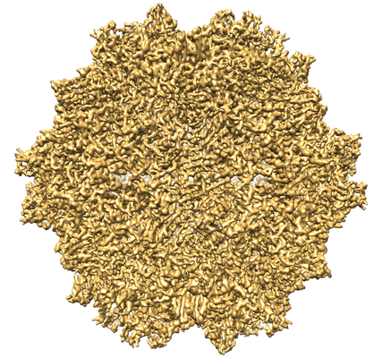 emd_8100.png
 http://ftp.pdbj.org/pub/emdb/structures/EMD-8100


 試料の構成要素
 Homo sapiens (ヒト)
 解析
解析 電子顕微鏡法
 FIELD EMISSION GUN
