 Movie
Movie Controller

[English] 日本語
 Yorodumi
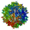
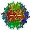
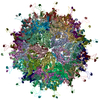
- PDB-5ipk: Structure of the R432A variant of Adeno-associated virus type 2 VLP -
+ Open data

- Basic information
| Entry | Database: PDB / ID: 5ipk | ||||||||||||||||||||||||||||||||||||||||||||||||||||||||||||
|---|---|---|---|---|---|---|---|---|---|---|---|---|---|---|---|---|---|---|---|---|---|---|---|---|---|---|---|---|---|---|---|---|---|---|---|---|---|---|---|---|---|---|---|---|---|---|---|---|---|---|---|---|---|---|---|---|---|---|---|---|---|
| Title | Structure of the R432A variant of Adeno-associated virus type 2 VLP | ||||||||||||||||||||||||||||||||||||||||||||||||||||||||||||
 Components Components | Capsid protein VP1 | ||||||||||||||||||||||||||||||||||||||||||||||||||||||||||||
 Keywords Keywords | VIRUS LIKE PARTICLE / Adeno-associated virus / R432A / gene therapy / icosahedral / dependoparvovirus | ||||||||||||||||||||||||||||||||||||||||||||||||||||||||||||
| Function / homology |  Function and homology information Function and homology informationsymbiont entry into host cell via permeabilization of host membrane / host cell nucleolus / T=1 icosahedral viral capsid / clathrin-dependent endocytosis of virus by host cell / virion attachment to host cell / structural molecule activity Similarity search - Function | ||||||||||||||||||||||||||||||||||||||||||||||||||||||||||||
| Biological species |  Adeno-associated virus - 2 Adeno-associated virus - 2 | ||||||||||||||||||||||||||||||||||||||||||||||||||||||||||||
| Method | ELECTRON MICROSCOPY / single particle reconstruction / cryo EM / Resolution: 3.7 Å | ||||||||||||||||||||||||||||||||||||||||||||||||||||||||||||
 Authors Authors | Drouin, L.M. / Lins, B. / Janssen, M.E. / Bennet, A. / Chipman, P. / McKenna, R. / Chen, W. / Muzyczka, N. / Cardone, G. / Baker, T.S. / Agbandje-McKenna, M. | ||||||||||||||||||||||||||||||||||||||||||||||||||||||||||||
| Funding support |  United States, 4items United States, 4items
| ||||||||||||||||||||||||||||||||||||||||||||||||||||||||||||
 Citation Citation | Journal: J Virol / Year: 2016 Title: Cryo-electron Microscopy Reconstruction and Stability Studies of the Wild Type and the R432A Variant of Adeno-associated Virus Type 2 Reveal that Capsid Structural Stability Is a Major Factor in Genome Packaging. Authors: Lauren M Drouin / Bridget Lins / Maria Janssen / Antonette Bennett / Paul Chipman / Robert McKenna / Weijun Chen / Nicholas Muzyczka / Giovanni Cardone / Timothy S Baker / Mavis Agbandje-McKenna / Abstract: The adeno-associated viruses (AAV) are promising therapeutic gene delivery vectors and better understanding of their capsid assembly and genome packaging mechanism is needed for improved vector ...The adeno-associated viruses (AAV) are promising therapeutic gene delivery vectors and better understanding of their capsid assembly and genome packaging mechanism is needed for improved vector production. Empty AAV capsids assemble in the nucleus prior to genome packaging by virally encoded Rep proteins. To elucidate the capsid determinants of this process, structural differences between wild-type (wt) AAV2 and a packaging deficient variant, AAV2-R432A, were examined using cryo-electron microscopy and three-dimensional image reconstruction both at an ∼5.0-Å resolution (medium) and also at 3.8- and 3.7-Å resolutions (high), respectively. The high resolution structures showed that removal of the arginine side chain in AAV2-R432A eliminated hydrogen bonding interactions, resulting in altered intramolecular and intermolecular interactions propagated from under the 3-fold axis toward the 5-fold channel. Consistent with these observations, differential scanning calorimetry showed an ∼10°C decrease in thermal stability for AAV2-R432A compared to wt-AAV2. In addition, the medium resolution structures revealed differences in the juxtaposition of the less ordered, N-terminal region of their capsid proteins, VP1/2/3. A structural rearrangement in AAV2-R432A repositioned the βA strand region under the icosahedral 2-fold axis rather than antiparallel to the βB strand, eliminating many intramolecular interactions. Thus, a single amino acid substitution can significantly alter the AAV capsid integrity to the extent of reducing its stability and possibly rendering it unable to tolerate the stress of genome packaging. Furthermore, the data show that the 2-, 3-, and 5-fold regions of the capsid contributed to producing the packaging defect and highlight a tight connection between the entire capsid in maintaining packaging efficiency. IMPORTANCE: The mechanism of AAV genome packaging is still poorly understood, particularly with respect to the capsid determinants of the required capsid-Rep interaction. Understanding this mechanism ...IMPORTANCE: The mechanism of AAV genome packaging is still poorly understood, particularly with respect to the capsid determinants of the required capsid-Rep interaction. Understanding this mechanism may aid in the improvement of AAV packaging efficiency, which is currently ∼1:10 (10%) genome packaged to empty capsid in vector preparations. This report identifies regions of the AAV capsid that play roles in genome packaging and that may be important for Rep recognition. It also demonstrates the need to maintain capsid stability for the success of this process. This information is important for efforts to improve AAV genome packaging and will also inform the engineering of AAV capsid variants for improved tropism, specific tissue targeting, and host antibody escape by defining amino acids that cannot be altered without detriment to infectious vector production. | ||||||||||||||||||||||||||||||||||||||||||||||||||||||||||||
| History |
|
- Structure visualization
| Movie |
Movie viewer |
|---|---|
| Structure viewer | Molecule: MolmilJmol/JSmol |
- Downloads & links
-Download
| PDBx/mmCIF format | 5ipk.cif.gz | 5.6 MB | Display | PDBx/mmCIF format |
|---|---|---|---|---|
| PDB format | pdb5ipk.ent.gz | Display | PDB format | |
| PDBx/mmJSON format | 5ipk.json.gz | Tree view | PDBx/mmJSON format | |
| Others |  Other downloads Other downloads |
-Validation report
| Arichive directory | https://data.pdbj.org/pub/pdb/validation_reports/ip/5ipkftp://data.pdbj.org/pub/pdb/validation_reports/ip/5ipk | HTTPS FTP |
|---|
-Related structure data
| Related structure data |  8100MC  8099C  5ipiC M: map data used to model this data C: citing same article ( |
|---|---|
| Similar structure data |


-Links
 PDBj
PDBj


- Assembly
| Deposited unit | 
|
|---|---|
| 1 |
|
-Components
| #1: Protein | Mass: 81945.234 Da / Num. of mol.: 60 / Mutation: R432A Source method: isolated from a genetically manipulated source Source: (gene. exp.) Adeno-associated virus - 2 / Gene: VP1 / Production host: unidentified baculovirus / References: UniProt: P03135Has protein modification | N | |
|---|
-Experimental details
-Experiment
| Experiment | Method: ELECTRON MICROSCOPY |
|---|---|
| EM experiment | Aggregation state: PARTICLE / 3D reconstruction method: single particle reconstruction |
- Sample preparation
| Component | Name: Adeno-associated virus - 2 / Type: VIRUS / Entity ID: all / Source: RECOMBINANT | ||||||||||||||||||||
|---|---|---|---|---|---|---|---|---|---|---|---|---|---|---|---|---|---|---|---|---|---|
| Source (natural) | Organism: Adeno-associated virus - 2 | ||||||||||||||||||||
| Source (recombinant) | Organism: unidentified baculovirus / Plasmid: pFBDVPm11 | ||||||||||||||||||||
| Details of virus | Empty: NO / Enveloped: NO / Isolate: STRAIN / Type: VIRUS-LIKE PARTICLE | ||||||||||||||||||||
| Natural host | Organism: Homo sapiens | ||||||||||||||||||||
| Buffer solution | pH: 7.4 | ||||||||||||||||||||
| Buffer component |
| ||||||||||||||||||||
| Specimen | Conc.: 1 mg/ml / Embedding applied: NO / Shadowing applied: NO / Staining applied: NO / Vitrification applied: YES | ||||||||||||||||||||
| Specimen support | Grid material: COPPER / Grid mesh size: 200 divisions/in. / Grid type: Quantifoil R2/2 | ||||||||||||||||||||
| Vitrification | Instrument: HOMEMADE PLUNGER / Cryogen name: ETHANE Details: Grid was blotted for 5 seconds before plunge freezing. |
- Electron microscopy imaging
| Experimental equipment |  Model: Tecnai Polara / Image courtesy: FEI Company |
|---|---|
| Microscopy | Model: FEI POLARA 300 |
| Electron gun | Electron source:  FIELD EMISSION GUN / Accelerating voltage: 300 kV / Illumination mode: FLOOD BEAM FIELD EMISSION GUN / Accelerating voltage: 300 kV / Illumination mode: FLOOD BEAM |
| Electron lens | Mode: BRIGHT FIELD / Nominal magnification: 59000 X / Calibrated magnification: 56924 X / Nominal defocus max: 3200 nm / Nominal defocus min: 1200 nm / Cs: 2.26 mm / C2 aperture diameter: 70 µm / Alignment procedure: COMA FREE |
| Specimen holder | Cryogen: NITROGEN / Specimen holder model: OTHER / Temperature (max): 97 K / Temperature (min): 90 K |
| Image recording | Average exposure time: 0.8 sec. / Electron dose: 20 e/Å2 / Film or detector model: KODAK SO-163 FILM / Num. of grids imaged: 1 / Num. of real images: 49 |
| Image scans | Sampling size: 6.35 µm |
- Processing
| Software | Name: PHENIX / Version: 1.10pre_2097: / Classification: refinement | ||||||||||||||||||||||||
|---|---|---|---|---|---|---|---|---|---|---|---|---|---|---|---|---|---|---|---|---|---|---|---|---|---|
| EM software |
| ||||||||||||||||||||||||
| CTF correction | Type: PHASE FLIPPING ONLY | ||||||||||||||||||||||||
| Particle selection | Num. of particles selected: 19457 | ||||||||||||||||||||||||
| 3D reconstruction | Resolution: 3.7 Å / Resolution method: FSC 0.5 CUT-OFF / Num. of particles: 19457 / Symmetry type: POINT | ||||||||||||||||||||||||
| Refinement | Highest resolution: 3.7 Å | ||||||||||||||||||||||||
| Refine LS restraints |
|
