Movie
Movie Controller


+ Open data

- Basic information
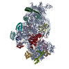
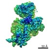
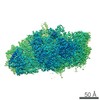
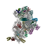
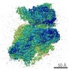
| Entry | Database: EMDB / ID: EMD-6780 | |||||||||
|---|---|---|---|---|---|---|---|---|---|---|
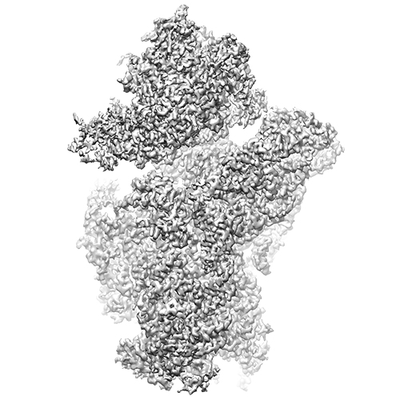
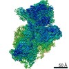
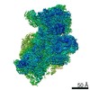
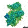
| Title | Small subunit of Toxoplasma gondii ribosome | |||||||||
 Map data Map data | ||||||||||
 Sample Sample |
| |||||||||
 Keywords Keywords | Toxoplasma gondii ribosome / rRNA / rprotein / RIBOSOME | |||||||||
| Function / homology |  Function and homology information Function and homology information90S preribosome / maturation of SSU-rRNA from tricistronic rRNA transcript (SSU-rRNA, 5.8S rRNA, LSU-rRNA) / maturation of SSU-rRNA / small-subunit processome / rRNA processing / ribosomal small subunit assembly / ribosomal small subunit biogenesis / small ribosomal subunit / small ribosomal subunit rRNA binding / cytosolic small ribosomal subunit ...90S preribosome / maturation of SSU-rRNA from tricistronic rRNA transcript (SSU-rRNA, 5.8S rRNA, LSU-rRNA) / maturation of SSU-rRNA / small-subunit processome / rRNA processing / ribosomal small subunit assembly / ribosomal small subunit biogenesis / small ribosomal subunit / small ribosomal subunit rRNA binding / cytosolic small ribosomal subunit / rRNA binding / structural constituent of ribosome / ribosome / translation / ribonucleoprotein complex / mRNA binding / nucleolus / RNA binding / zinc ion binding / membrane / cytoplasm / cytosol Similarity search - Function | |||||||||
| Biological species |  | |||||||||
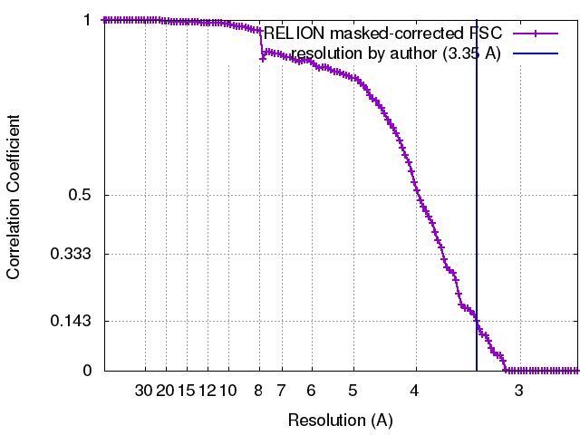
| Method | single particle reconstruction / cryo EM / Resolution: 3.35 Å | |||||||||
 Authors Authors | LI Z / Guo Q | |||||||||
 Citation Citation | Journal: Cell Res / Year: 2017 Title: Cryo-EM structures of the 80S ribosomes from human parasites Trichomonas vaginalis and Toxoplasma gondii. Authors: Zhifei Li / Qiang Guo / Lvqin Zheng / Yongsheng Ji / Yi-Ting Xie / De-Hua Lai / Zhao-Rong Lun / Xun Suo / Ning Gao /  Abstract: As an indispensable molecular machine universal in all living organisms, the ribosome has been selected by evolution to be the natural target of many antibiotics and small-molecule inhibitors. High- ...As an indispensable molecular machine universal in all living organisms, the ribosome has been selected by evolution to be the natural target of many antibiotics and small-molecule inhibitors. High-resolution structures of pathogen ribosomes are crucial for understanding the general and unique aspects of translation control in disease-causing microbes. With cryo-electron microscopy technique, we have determined structures of the cytosolic ribosomes from two human parasites, Trichomonas vaginalis and Toxoplasma gondii, at resolution of 3.2-3.4 Å. Although the ribosomal proteins from both pathogens are typical members of eukaryotic families, with a co-evolution pattern between certain species-specific insertions/extensions and neighboring ribosomal RNA (rRNA) expansion segments, the sizes of their rRNAs are sharply different. Very interestingly, rRNAs of T. vaginalis are in size comparable to prokaryotic counterparts, with nearly all the eukaryote-specific rRNA expansion segments missing. These structures facilitate the dissection of evolution path for ribosomal proteins and RNAs, and may aid in design of novel translation inhibitors. | |||||||||
| History |
|
- Structure visualization
| Movie |
Movie viewer |
|---|---|
| Structure viewer | EM map: SurfViewMolmilJmol/JSmol |
| Supplemental images |
- Downloads & links
-EMDB archive
| Map data | emd_6780.map.gz | 9.1 MB | EMDB map data format | |
|---|---|---|---|---|
| Header (meta data) | emd-6780-v30.xmlemd-6780.xml | 40.3 KB 40.3 KB | Display Display | EMDB header |
| FSC (resolution estimation) | emd_6780_fsc.xml | 11.1 KB | Display | FSC data file |
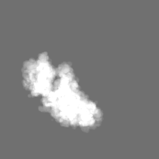
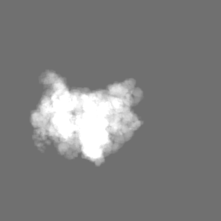
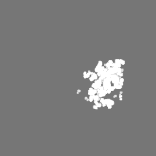
| Images |  emd_6780.png emd_6780.png | 76.9 KB | ||
| Masks | emd_6780_msk_1.map | 125 MB | Mask map | |
| Filedesc metadata | emd-6780.cif.gz | 9.8 KB | ||
| Archive directory |  http://ftp.pdbj.org/pub/emdb/structures/EMD-6780ftp://ftp.pdbj.org/pub/emdb/structures/EMD-6780 http://ftp.pdbj.org/pub/emdb/structures/EMD-6780ftp://ftp.pdbj.org/pub/emdb/structures/EMD-6780 | HTTPS FTP |
-Related structure data
| Related structure data |  5xxuMC  6778C 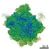 6784C 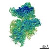 6788C 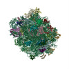 5xxbC 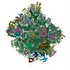 5xy3C 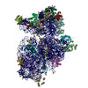 5xyiC C: citing same article ( M: atomic model generated by this map |
|---|---|
| Similar structure data |
-Links
| EMDB pages | EMDB (EBI/PDBe) / EMDataResource |
|---|---|
| Related items in Molecule of the Month |


-Map
| File | Download / File: emd_6780.map.gz / Format: CCP4 / Size: 125 MB / Type: IMAGE STORED AS FLOATING POINT NUMBER (4 BYTES) | ||||||||||||||||||||||||||||||||||||||||||||||||||||||||||||
|---|---|---|---|---|---|---|---|---|---|---|---|---|---|---|---|---|---|---|---|---|---|---|---|---|---|---|---|---|---|---|---|---|---|---|---|---|---|---|---|---|---|---|---|---|---|---|---|---|---|---|---|---|---|---|---|---|---|---|---|---|---|
| Projections & slices | Image control
Images are generated by Spider. | ||||||||||||||||||||||||||||||||||||||||||||||||||||||||||||
| Voxel size | X=Y=Z: 1.32 Å | ||||||||||||||||||||||||||||||||||||||||||||||||||||||||||||
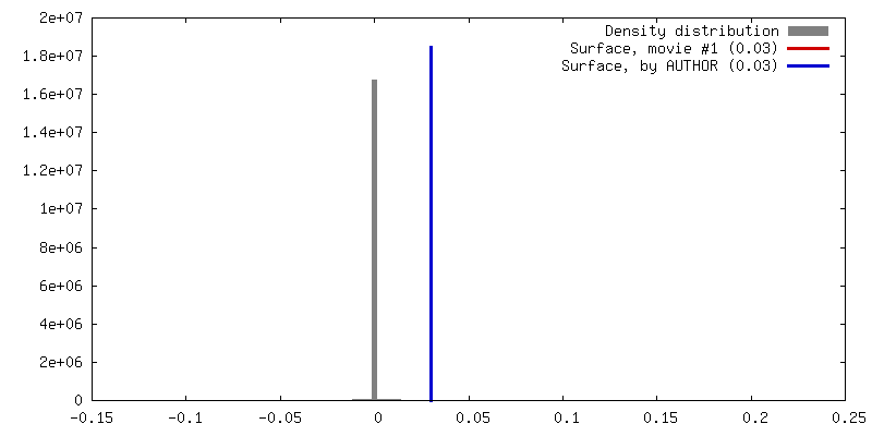
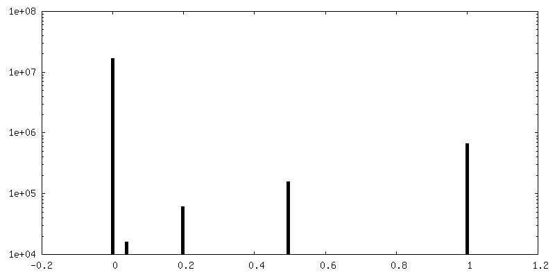
| Density |
| ||||||||||||||||||||||||||||||||||||||||||||||||||||||||||||
| Symmetry | Space group: 1 | ||||||||||||||||||||||||||||||||||||||||||||||||||||||||||||
| Details | EMDB XML:
CCP4 map header:
| ||||||||||||||||||||||||||||||||||||||||||||||||||||||||||||
 Z (Sec.)
 Y (Row.)
 X (Col.)


-Supplemental data
-Mask #1
| File | emd_6780_msk_1.map | ||||||||||||
|---|---|---|---|---|---|---|---|---|---|---|---|---|---|
| Projections & Slices |
| ||||||||||||
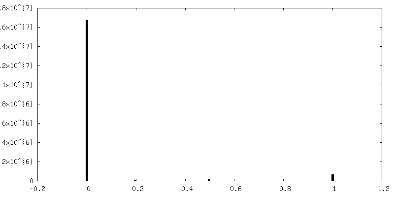
| Density Histograms |

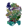
- Sample components
+Entire : Small subunit of Toxoplasma gondii ribosome
+Supramolecule #1: Small subunit of Toxoplasma gondii ribosome
+Macromolecule #1: 18S RNA
+Macromolecule #2: Ribosomal protein uS2
+Macromolecule #3: Ribosomal protein eS1
+Macromolecule #4: Ribosomal protein uS5
+Macromolecule #5: Ribosomal protein uS3
+Macromolecule #6: Ribosomal protein eS4
+Macromolecule #7: Ribosomal protein uS7
+Macromolecule #8: Ribosomal protein eS6
+Macromolecule #9: Ribosomal protein eS7
+Macromolecule #10: Ribosomal protein eS8
+Macromolecule #11: Ribosomal protein uS4
+Macromolecule #12: Ribosomal protein eS10
+Macromolecule #13: Ribosomal protein uS17
+Macromolecule #14: Ribosomal protein eS12
+Macromolecule #15: Ribosomal protein uS15
+Macromolecule #16: Ribosomal protein uS11
+Macromolecule #17: Ribosomal protein uS19
+Macromolecule #18: Ribosomal protein uS9
+Macromolecule #19: Ribosomal protein eS17
+Macromolecule #20: Ribosomal protein uS13
+Macromolecule #21: Ribosomal protein eS19
+Macromolecule #22: Ribosomal protein uS10
+Macromolecule #23: Ribosomal protein eS21
+Macromolecule #24: Ribosomal protein uS8
+Macromolecule #25: Ribosomal protein uS12
+Macromolecule #26: Ribosomal protein eS24
+Macromolecule #27: Ribosomal protein eS25
+Macromolecule #28: Ribosomal protein eS26
+Macromolecule #29: Ribosomal protein eS27
+Macromolecule #30: Ribosomal protein eS28
+Macromolecule #31: Ribosomal protein uS14
+Macromolecule #32: Ribosomal protein eS30
+Macromolecule #33: Ribosomal protein eS31
+Macromolecule #34: Ribosomal protein eL41
-Experimental details
-Structure determination
| Method | cryo EM |
|---|---|
 Processing Processing | single particle reconstruction |
| Aggregation state | particle |
-Sample preparation
| Buffer | pH: 7.4 |
|---|---|
| Vitrification | Cryogen name: ETHANE |
- Electron microscopy
| Microscope | FEI TITAN KRIOS |
|---|---|
| Image recording | Film or detector model: GATAN K2 SUMMIT (4k x 4k) / Average electron dose: 2.0 e/Å2 |
| Electron beam | Acceleration voltage: 300 kV / Electron source:  FIELD EMISSION GUN FIELD EMISSION GUN |
| Electron optics | Illumination mode: FLOOD BEAM / Imaging mode: BRIGHT FIELD |
| Experimental equipment |  Model: Titan Krios / Image courtesy: FEI Company |