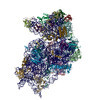
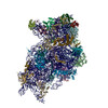
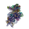
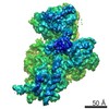
- EMDB-11520: Cryo-EM structure of a late human pre-40S ribosomal subunit - State H1 -
+
Open data
ID or keywords:
Loading...
-
Basic information
Entry
Database: EMDB / ID: EMD-11520
Title
Cryo-EM structure of a late human pre-40S ribosomal subunit - State H1
Map data
Sample
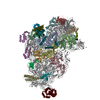
Complex: Cryo-EM structure of a late human pre-40S ribosomal subunit - State H1
RNA: x 1 types
Protein or peptide: x 36 types
Ligand: x 4 types
Keywords
Ribosome Biogenesis / Pre-40S / RIBOSOME
Function / homology
Function and homology information
methyltransferase complex / phenylalanyl-tRNA aminoacylation / phenylalanine-tRNA ligase activity / positive regulation of rRNA processing / negative regulation of endoplasmic reticulum unfolded protein response / oxidized pyrimidine DNA binding / response to TNF agonist / positive regulation of base-excision repair / positive regulation of respiratory burst involved in inflammatory response / positive regulation of gastrulation ...methyltransferase complex / phenylalanyl-tRNA aminoacylation / phenylalanine-tRNA ligase activity / positive regulation of rRNA processing / negative regulation of endoplasmic reticulum unfolded protein response / oxidized pyrimidine DNA binding / response to TNF agonist / positive regulation of base-excision repair / positive regulation of respiratory burst involved in inflammatory response / positive regulation of gastrulation / protein tyrosine kinase inhibitor activity / positive regulation of intrinsic apoptotic signaling pathway in response to DNA damage / positive regulation of ubiquitin-protein transferase activity / IRE1-RACK1-PP2A complex / positive regulation of DNA-templated transcription initiation / positive regulation of Golgi to plasma membrane protein transport / nucleolus organization / TNFR1-mediated ceramide production / negative regulation of RNA splicing / neural crest cell differentiation / supercoiled DNA binding / NF-kappaB complex / negative regulation of DNA repair / cytoplasmic translational initiation / cysteine-type endopeptidase activator activity involved in apoptotic process / preribosome, small subunit precursor / oxidized purine DNA binding / rRNA modification in the nucleus and cytosol / negative regulation of intrinsic apoptotic signaling pathway in response to hydrogen peroxide / regulation of establishment of cell polarity / negative regulation of bicellular tight junction assembly / ubiquitin-like protein conjugating enzyme binding / negative regulation of phagocytosis / erythrocyte homeostasis / cytoplasmic side of rough endoplasmic reticulum membrane / Formation of the ternary complex, and subsequently, the 43S complex / ion channel inhibitor activity / protein kinase A binding / laminin receptor activity / pigmentation / Ribosomal scanning and start codon recognition / positive regulation of mitochondrial depolarization / Translation initiation complex formation / negative regulation of Wnt signaling pathway / fibroblast growth factor binding / Protein hydroxylation / monocyte chemotaxis / BH3 domain binding / negative regulation of translational frameshifting / regulation of adenylate cyclase-activating G protein-coupled receptor signaling pathway / TOR signaling / mTORC1-mediated signalling / SARS-CoV-1 modulates host translation machinery / iron-sulfur cluster binding / positive regulation of GTPase activity / regulation of cell division / cellular response to ethanol / Peptide chain elongation / Selenocysteine synthesis / Formation of a pool of free 40S subunits / negative regulation of protein binding / Eukaryotic Translation Termination / positive regulation of intrinsic apoptotic signaling pathway by p53 class mediator / protein serine/threonine kinase inhibitor activity / SRP-dependent cotranslational protein targeting to membrane / Response of EIF2AK4 (GCN2) to amino acid deficiency / negative regulation of respiratory burst involved in inflammatory response / ubiquitin ligase inhibitor activity / Viral mRNA Translation / endonucleolytic cleavage to generate mature 3'-end of SSU-rRNA from (SSU-rRNA, 5.8S rRNA, LSU-rRNA) / negative regulation of ubiquitin-dependent protein catabolic process / Nonsense Mediated Decay (NMD) independent of the Exon Junction Complex (EJC) / positive regulation of signal transduction by p53 class mediator / GTP hydrolysis and joining of the 60S ribosomal subunit / L13a-mediated translational silencing of Ceruloplasmin expression / Hydrolases; Acting on acid anhydrides; In phosphorus-containing anhydrides / Major pathway of rRNA processing in the nucleolus and cytosol / regulation of translational fidelity / positive regulation of microtubule polymerization / phagocytic cup / Nonsense Mediated Decay (NMD) enhanced by the Exon Junction Complex (EJC) / spindle assembly / positive regulation of intrinsic apoptotic signaling pathway / Protein methylation / endonucleolytic cleavage in ITS1 to separate SSU-rRNA from 5.8S rRNA and LSU-rRNA from tricistronic rRNA transcript (SSU-rRNA, 5.8S rRNA, LSU-rRNA) / translation regulator activity / Nuclear events stimulated by ALK signaling in cancer / rough endoplasmic reticulum / positive regulation of cell cycle / laminin binding / ribosomal small subunit export from nucleus / Amplification of signal from unattached kinetochores via a MAD2 inhibitory signal / DNA-(apurinic or apyrimidinic site) endonuclease activity / translation initiation factor activity / gastrulation / signaling adaptor activity / Maturation of protein E / translation initiation factor binding / negative regulation of protein ubiquitination / Maturation of protein E Similarity search - Function
Probable RNA-binding protein EIF1AD / Serine/threonine-protein kinase Rio1 / : / RIO kinase, conserved site / RIO1/ZK632.3/MJ0444 family signature. / Phenylalanine-tRNA ligase, class IIc, beta subunit / B3/B4 tRNA-binding domain / Phenylalanyl-tRNA synthetase-like, B3/B4 / B3/4 domain / RIO kinase ...Probable RNA-binding protein EIF1AD / Serine/threonine-protein kinase Rio1 / : / RIO kinase, conserved site / RIO1/ZK632.3/MJ0444 family signature. / Phenylalanine-tRNA ligase, class IIc, beta subunit / B3/B4 tRNA-binding domain / Phenylalanyl-tRNA synthetase-like, B3/B4 / B3/4 domain / RIO kinase / RIO-like kinase / : / RIO domain / eukaryotic translation initiation factor 1A / Translation initiation factor 1A (eIF-1A) / Leucine rich repeat 4 / Leucine Rich repeats (2 copies) / RNA-binding domain, S1, IF1 type / Translation initiation factor 1A / IF-1 / S1 domain IF1 type profile. / Leucine-rich repeats, bacterial type / 40S ribosomal protein SA / 40S ribosomal protein SA, C-terminal domain / 40S ribosomal protein SA C-terminus / Ubiquitin-like protein FUBI / Leucine rich repeat / Leucine-rich repeat, typical subtype / Leucine-rich repeats, typical (most populated) subfamily / : / Ribosomal protein S26e signature. / Ribosomal protein S21e, conserved site / Ribosomal protein S21e signature. / : / Ribosomal protein S12e signature. / Ribosomal protein S26e / Ribosomal protein S26e superfamily / Ribosomal protein S26e / Ribosomal protein S12e / Small (40S) ribosomal subunit Asc1/RACK1 / Ribosomal protein S5, eukaryotic/archaeal / Ribosomal protein S19e, conserved site / Ribosomal protein S19e signature. / Ribosomal protein S21e / Ribosomal protein S21e superfamily / Ribosomal protein S21e / Ribosomal protein S2, eukaryotic / 40S Ribosomal protein S10 / S27a-like superfamily / Plectin/S10, N-terminal / Plectin/S10 domain / Ribosomal protein S10, eukaryotic/archaeal / Ribosomal protein S30 / Ribosomal protein S30 / Ribosomal protein S25 / S25 ribosomal protein / Leucine-rich repeat profile. / Ribosomal protein S8e subdomain, eukaryotes / : / Ribosomal protein S17e, conserved site / Ribosomal protein S17e signature. / Ribosomal protein S7e signature. / Ribosomal protein S27a / Ribosomal protein S27a / Ribosomal protein S27a / Ribosomal protein S2, eukaryotic/archaeal / 40S ribosomal protein S29/30S ribosomal protein S14 type Z / Ribosomal protein S3, eukaryotic/archaeal / Ribosomal protein S3Ae, conserved site / Ribosomal protein S3Ae signature. / Ribosomal protein S8e, conserved site / Ribosomal protein S8e signature. / Ribosomal protein S27e signature. / 40S ribosomal protein S4, C-terminal domain / 40S ribosomal protein S4 C-terminus / Ribosomal protein S19A/S15e / Ribosomal protein S19e / Ribosomal protein S19e / Ribosomal_S19e / Ribosomal protein S4e, N-terminal, conserved site / Ribosomal protein S4e signature. / Ribosomal protein S17e / Ribosomal protein S17e-like superfamily / Ribosomal S17 / Ribosomal protein S6, eukaryotic / 40S ribosomal protein S1/3, eukaryotes / 40S ribosomal protein S11, N-terminal / Ribosomal_S17 N-terminal / Ribosomal protein S7e / Ribosomal protein S7e / : / Ribosomal S24e conserved site / Ribosomal protein S24e signature. / Ribosomal protein S4e, N-terminal / RS4NT (NUC023) domain / Ribosomal protein S4, KOW domain / Ribosomal protein S4e / Ribosomal protein S4e, central region / Ribosomal protein S4e, central domain superfamily / Ribosomal family S4e / Ribosomal protein S28e conserved site Similarity search - Domain/homology
Small ribosomal subunit protein eS17 / Small ribosomal subunit protein uS2 / Small ribosomal subunit protein uS5 / Small ribosomal subunit protein uS3 / Small ribosomal subunit protein eS12 / Small ribosomal subunit protein eS19 / Small ribosomal subunit protein eS27 / Small ribosomal subunit protein uS4 / Small ribosomal subunit protein uS7 / Small ribosomal subunit protein eS10 ...Small ribosomal subunit protein eS17 / Small ribosomal subunit protein uS2 / Small ribosomal subunit protein uS5 / Small ribosomal subunit protein uS3 / Small ribosomal subunit protein eS12 / Small ribosomal subunit protein eS19 / Small ribosomal subunit protein eS27 / Small ribosomal subunit protein uS4 / Small ribosomal subunit protein uS7 / Small ribosomal subunit protein eS10 / Small ribosomal subunit protein uS10 / Small ribosomal subunit protein eS1 / Small ribosomal subunit protein eS7 / Small ribosomal subunit protein eS8 / Small ribosomal subunit protein uS8 / Small ribosomal subunit protein uS9 / Small ribosomal subunit protein uS11 / Small ribosomal subunit protein uS12 / Small ribosomal subunit protein uS13 / Small ribosomal subunit protein uS14 / Small ribosomal subunit protein uS15 / Small ribosomal subunit protein uS17 / Small ribosomal subunit protein eS4, X isoform / Small ribosomal subunit protein eS6 / Small ribosomal subunit protein uS19 / Small ribosomal subunit protein eS24 / Small ribosomal subunit protein eS25 / Small ribosomal subunit protein eS26 / Small ribosomal subunit protein eS28 / Ubiquitin-like FUBI-ribosomal protein eS30 fusion protein / Ubiquitin-ribosomal protein eS31 fusion protein / Small ribosomal subunit protein eS21 / Small ribosomal subunit protein RACK1 / Leucine-rich repeat-containing protein 47 / Probable RNA-binding protein EIF1AD / Serine/threonine-protein kinase RIO1 Similarity search - Component
Biological species
Homo sapiens (human)
Method
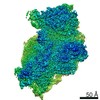
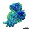
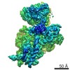
single particle reconstruction / cryo EM / Resolution: 2.6 Å
Journal: Nature / Year: 2020 Title: Structural basis for the final steps of human 40S ribosome maturation. Authors: Michael Ameismeier / Ivo Zemp / Jasmin van den Heuvel / Matthias Thoms / Otto Berninghausen / Ulrike Kutay / Roland Beckmann / Abstract: Eukaryotic ribosomes consist of a small 40S and a large 60S subunit that are assembled in a highly coordinated manner. More than 200 factors ensure correct modification, processing and folding of ...Eukaryotic ribosomes consist of a small 40S and a large 60S subunit that are assembled in a highly coordinated manner. More than 200 factors ensure correct modification, processing and folding of ribosomal RNA and the timely incorporation of ribosomal proteins. Small subunit maturation ends in the cytosol, when the final rRNA precursor, 18S-E, is cleaved at site 3 by the endonuclease NOB1. Previous structures of human 40S precursors have shown that NOB1 is kept in an inactive state by its partner PNO1. The final maturation events, including the activation of NOB1 for the decisive rRNA-cleavage step and the mechanisms driving the dissociation of the last biogenesis factors have, however, remained unresolved. Here we report five cryo-electron microscopy structures of human 40S subunit precursors, which describe the compositional and conformational progression during the final steps of 40S assembly. Our structures explain the central role of RIOK1 in the displacement and dissociation of PNO1, which in turn allows conformational changes and activation of the endonuclease NOB1. In addition, we observe two factors, eukaryotic translation initiation factor 1A domain-containing protein (EIF1AD) and leucine-rich repeat-containing protein 47 (LRRC47), which bind to late pre-40S particles near RIOK1 and the central rRNA helix 44. Finally, functional data shows that EIF1AD is required for efficient assembly factor recycling and 18S-E processing. Our results thus enable a detailed understanding of the last steps in 40S formation in human cells and, in addition, provide evidence for principal differences in small ribosomal subunit formation between humans and the model organism Saccharomyces cerevisiae.
History
Deposition
Jul 29, 2020
-
Header (metadata) release
Dec 2, 2020
-
Map release
Dec 2, 2020
-
Update
Jun 24, 2026
-
Current status
Jun 24, 2026
Processing site: PDBe / Status: Released
-
Structure visualization
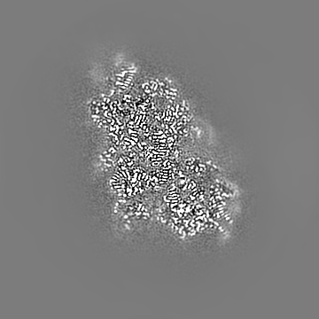
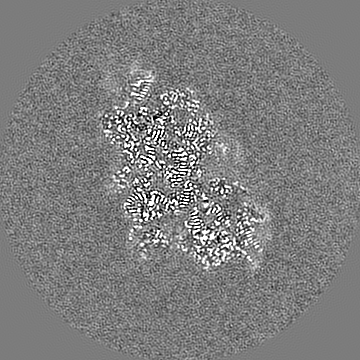
Movie
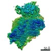
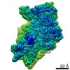
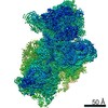
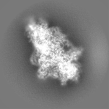
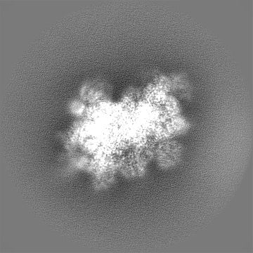
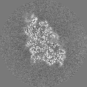
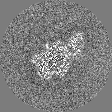
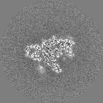
Surface view with section colored by density value
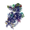
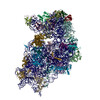
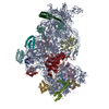
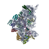
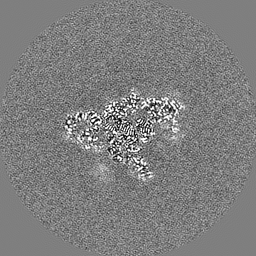
Entire : Cryo-EM structure of a late human pre-40S ribosomal subunit - State H1
Entire
Name: Cryo-EM structure of a late human pre-40S ribosomal subunit - State H1
Components
Complex: Cryo-EM structure of a late human pre-40S ribosomal subunit - State H1
RNA: pre-18S ribosomal RNA
Protein or peptide: 40S ribosomal protein SA
Protein or peptide: 40S ribosomal protein S3a
Protein or peptide: 40S ribosomal protein S2
Protein or peptide: 40S ribosomal protein S26
Protein or peptide: 40S ribosomal protein S4, X isoform
Protein or peptide: 40S ribosomal protein S3
Protein or peptide: 40S ribosomal protein S6
Protein or peptide: 40S ribosomal protein S7
Protein or peptide: 40S ribosomal protein S8
Protein or peptide: 40S ribosomal protein S9
Protein or peptide: 40S ribosomal protein S5
Protein or peptide: 40S ribosomal protein S11
Protein or peptide: 40S ribosomal protein S10
Protein or peptide: 40S ribosomal protein S13
Protein or peptide: 40S ribosomal protein S14
Protein or peptide: 40S ribosomal protein S12
Protein or peptide: 40S ribosomal protein S15
Protein or peptide: 40S ribosomal protein S17
Protein or peptide: 40S ribosomal protein S16
Protein or peptide: 40S ribosomal protein S18
Protein or peptide: 40S ribosomal protein S19
Protein or peptide: 40S ribosomal protein S21
Protein or peptide: 40S ribosomal protein S15a
Protein or peptide: 40S ribosomal protein S23
Protein or peptide: 40S ribosomal protein S24
Protein or peptide: 40S ribosomal protein S20
Protein or peptide: 40S ribosomal protein S25
Protein or peptide: 40S ribosomal protein S27
Protein or peptide: 40S ribosomal protein S28
Protein or peptide: 40S ribosomal protein S29
Protein or peptide: 40S ribosomal protein S30
Protein or peptide: Ubiquitin-40S ribosomal protein S27a
Protein or peptide: Receptor of activated protein C kinase 1
Protein or peptide: Probable RNA-binding protein EIF1AD
Protein or peptide: Leucine-rich repeat-containing protein 47
Protein or peptide: Serine/threonine-protein kinase RIO1
Ligand: MAGNESIUM ION
Ligand: ZINC ION
Ligand: ASPARTIC ACID
Ligand: ADENOSINE-5'-TRIPHOSPHATE
+
Supramolecule #1: Cryo-EM structure of a late human pre-40S ribosomal subunit - State H1
Supramolecule
Name: Cryo-EM structure of a late human pre-40S ribosomal subunit - State H1 type: complex / ID: 1 / Parent: 0 / Macromolecule list: #1-#37 / Details: Map filtered at local resolution
Source (natural)
Organism: Homo sapiens (human) / Strain: HEK293T
+
Macromolecule #1: pre-18S ribosomal RNA
Macromolecule
Name: pre-18S ribosomal RNA / type: rna / ID: 1 / Number of copies: 1
Source (natural)
Organism: Homo sapiens (human)
Molecular weight
Theoretical: 603.524062 KDa
Sequence
String: UACCUGGUUG AUCCUGCCAG UAGCAU(A2M)UGC UUG(PSU)C(PSU)CAAA GAUUAAGCCA UGCAUGUCUA AGUACGCA C GGCCGGUACA GUGAAACUGC GAA(PSU)GGCUC(A2M) UUAAA(PSU)CAG(PSU) UAUGGU(OMU)CC(PSU) U (OMU)GGUCGCU ...String:
In the structure databanks used in Yorodumi, some data are registered as the other names, "COVID-19 virus" and "2019-nCoV". Here are the details of the virus and the list of structure data.
Jan 31, 2019. EMDB accession codes are about to change! (news from PDBe EMDB page)
EMDB accession codes are about to change! (news from PDBe EMDB page)
The allocation of 4 digits for EMDB accession codes will soon come to an end. Whilst these codes will remain in use, new EMDB accession codes will include an additional digit and will expand incrementally as the available range of codes is exhausted. The current 4-digit format prefixed with “EMD-” (i.e. EMD-XXXX) will advance to a 5-digit format (i.e. EMD-XXXXX), and so on. It is currently estimated that the 4-digit codes will be depleted around Spring 2019, at which point the 5-digit format will come into force.
The EM Navigator/Yorodumi systems omit the EMD- prefix.
Related info.:Q: What is EMD? / ID/Accession-code notation in Yorodumi/EM Navigator
Yorodumi is a browser for structure data from EMDB, PDB, SASBDB, etc.
This page is also the successor to EM Navigator detail page, and also detail information page/front-end page for Omokage search.
The word "yorodu" (or yorozu) is an old Japanese word meaning "ten thousand". "mi" (miru) is to see.
Related info.:EMDB / PDB / SASBDB / Comparison of 3 databanks / Yorodumi Search / Aug 31, 2016. New EM Navigator & Yorodumi / Yorodumi Papers / Jmol/JSmol / Function and homology information / Changes in new EM Navigator and Yorodumi
 Movie
Movie Controller

 Yorodumi
 Open data
 Basic information
 Map data
 Sample
 Keywords
 Function and homology information
 Homo sapiens (human)
 Authors
 Germany, 1 items
  Citation
 Structure visualization
 Downloads & links
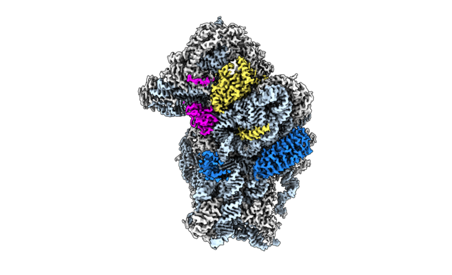 emd_11520.png
 http://ftp.pdbj.org/pub/emdb/structures/EMD-11520
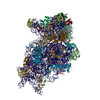


 Z (Sec.)
 Y (Row.)
 X (Col.)


 Sample components

 Processing
 Electron microscopy
 FIELD EMISSION GUN