 Movie
Movie Controller

[English] 日本語
 Yorodumi
- EMDB-3579: 30S ribosomal subunit with partial density for ribosomal protein S1 -
+ Open data

- Basic information
| Entry | Database: EMDB / ID: EMD-3579 | |||||||||
|---|---|---|---|---|---|---|---|---|---|---|
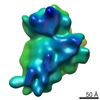
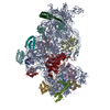
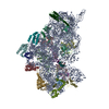
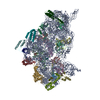
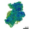
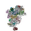
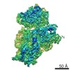
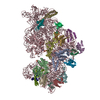
| Title | 30S ribosomal subunit with partial density for ribosomal protein S1 | |||||||||
 Map data Map data | None | |||||||||
 Sample Sample |
| |||||||||
| Biological species |  | |||||||||
| Method | single particle reconstruction / cryo EM / Resolution: 6.4 Å | |||||||||
 Authors Authors | Kohler R / Mooney RA / Mills DJ / Landick R / Cramer P | |||||||||
 Citation Citation | Journal: Science / Year: 2017 Title: Architecture of a transcribing-translating expressome. Authors: R Kohler / R A Mooney / D J Mills / R Landick / P Cramer /   Abstract: DNA transcription is functionally coupled to messenger RNA (mRNA) translation in bacteria, but how this is achieved remains unclear. Here we show that RNA polymerase (RNAP) and the ribosome of can ...DNA transcription is functionally coupled to messenger RNA (mRNA) translation in bacteria, but how this is achieved remains unclear. Here we show that RNA polymerase (RNAP) and the ribosome of can form a defined transcribing and translating "expressome" complex. The cryo-electron microscopic structure of the expressome reveals continuous protection of ~30 nucleotides of mRNA extending from the RNAP active center to the ribosome decoding center. The RNAP-ribosome interface includes the RNAP subunit α carboxyl-terminal domain, which is required for RNAP-ribosome interaction in vitro and for pronounced cell growth defects upon translation inhibition in vivo, consistent with its function in transcription-translation coupling. The expressome structure can only form during transcription elongation and explains how translation can prevent transcriptional pausing, backtracking, and termination. | |||||||||
| History |
|
- Structure visualization
| Movie |
 Movie viewer Movie viewer |
|---|---|
| Structure viewer | EM map: SurfViewMolmilJmol/JSmol |
| Supplemental images |

- Downloads & links
-EMDB archive
| Map data | emd_3579.map.gz | 3.9 MB | EMDB map data format | |
|---|---|---|---|---|
| Header (meta data) | emd-3579-v30.xmlemd-3579.xml | 8.9 KB 8.9 KB | Display Display | EMDB header |
| Images |  emd_3579.png emd_3579.png | 50.2 KB | ||
| Archive directory |  http://ftp.pdbj.org/pub/emdb/structures/EMD-3579ftp://ftp.pdbj.org/pub/emdb/structures/EMD-3579 http://ftp.pdbj.org/pub/emdb/structures/EMD-3579ftp://ftp.pdbj.org/pub/emdb/structures/EMD-3579 | HTTPS FTP |
-Related structure data


-Links
| EMDB pages | EMDB (EBI/PDBe) / EMDataResource |
|---|---|
| Related items in Molecule of the Month |

-Map
| File | Download / File: emd_3579.map.gz / Format: CCP4 / Size: 67 MB / Type: IMAGE STORED AS FLOATING POINT NUMBER (4 BYTES) | ||||||||||||||||||||||||||||||||||||||||||||||||||||||||||||
|---|---|---|---|---|---|---|---|---|---|---|---|---|---|---|---|---|---|---|---|---|---|---|---|---|---|---|---|---|---|---|---|---|---|---|---|---|---|---|---|---|---|---|---|---|---|---|---|---|---|---|---|---|---|---|---|---|---|---|---|---|---|
| Annotation | None | ||||||||||||||||||||||||||||||||||||||||||||||||||||||||||||
| Projections & slices | Image control
Images are generated by Spider. | ||||||||||||||||||||||||||||||||||||||||||||||||||||||||||||
| Voxel size | X=Y=Z: 1.86 Å | ||||||||||||||||||||||||||||||||||||||||||||||||||||||||||||
| Density |
| ||||||||||||||||||||||||||||||||||||||||||||||||||||||||||||
| Symmetry | Space group: 1 | ||||||||||||||||||||||||||||||||||||||||||||||||||||||||||||
| Details | EMDB XML:
CCP4 map header:
| ||||||||||||||||||||||||||||||||||||||||||||||||||||||||||||
 Z (Sec.)
 Y (Row.)
 X (Col.)


-Supplemental data
- Sample components
-Entire : the sample contained a mixture of in vitro assembled E.coli expre...
| Entire | Name: the sample contained a mixture of in vitro assembled E.coli expressomes and free ribosomes (see related entry EMDB 3580 and related publication). This structure shows additional density for ...Name: the sample contained a mixture of in vitro assembled E.coli expressomes and free ribosomes (see related entry EMDB 3580 and related publication). This structure shows additional density for ribosomal protein S1 and weak density for the RNA Polymerase. |
|---|---|
| Components |
|
-Supramolecule #1: the sample contained a mixture of in vitro assembled E.coli expre...
| Supramolecule | Name: the sample contained a mixture of in vitro assembled E.coli expressomes and free ribosomes (see related entry EMDB 3580 and related publication). This structure shows additional density for ...Name: the sample contained a mixture of in vitro assembled E.coli expressomes and free ribosomes (see related entry EMDB 3580 and related publication). This structure shows additional density for ribosomal protein S1 and weak density for the RNA Polymerase. type: complex / ID: 1 / Parent: 0 / Macromolecule list: all |
|---|---|
| Source (natural) | Organism: |
-Macromolecule #1: ribosome
| Macromolecule | Name: ribosome / type: other / ID: 1 / Classification: other |
|---|---|
| Source (natural) | Organism: |
| Sequence | String: not applic able |
-Experimental details
-Structure determination
| Method | cryo EM |
|---|---|
 Processing Processing | single particle reconstruction |
| Aggregation state | particle |
-Sample preparation
| Buffer | pH: 7.6 |
|---|---|
| Vitrification | Cryogen name: ETHANE |
- Electron microscopy
| Microscope | FEI POLARA 300 |
|---|---|
| Image recording | Film or detector model: GATAN K2 SUMMIT (4k x 4k) / Average electron dose: 16.0 e/Å2 |
| Electron beam | Acceleration voltage: 300 kV / Electron source:  FIELD EMISSION GUN FIELD EMISSION GUN |
| Electron optics | Illumination mode: SPOT SCAN / Imaging mode: BRIGHT FIELD |
| Experimental equipment |  Model: Tecnai Polara / Image courtesy: FEI Company |
-Image processing
| CTF correction | Software - Name: RELION (ver. 1.4) |
|---|---|
| Final reconstruction | Applied symmetry - Point group: C1 (asymmetric) / Resolution.type: BY AUTHOR / Resolution: 6.4 Å / Resolution method: FSC 0.143 CUT-OFF / Software - Name: RELION (ver. 1.4) / Number images used: 71835 |
| Initial angle assignment | Type: PROJECTION MATCHING / Software - Name: RELION |
| Final angle assignment | Type: PROJECTION MATCHING / Software - Name: RELION (ver. 1.4) |
