Movie
Movie Controller

[English] 日本語
 Yorodumi
- EMDB-3308: HIV-1 cleaved wild type JR-FL EnvdCT trimer in complex with PGT15... -
+ Open data

- Basic information
| Entry | Database: EMDB / ID: EMD-3308 | |||||||||
|---|---|---|---|---|---|---|---|---|---|---|
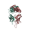
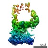
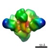
| Title | HIV-1 cleaved wild type JR-FL EnvdCT trimer in complex with PGT151 at 4.19 A resolution | |||||||||
 Map data Map data | Wild type JR-FL EnvdCT in complex with PGT151, with Fab constant region and detergent micelle masked out. | |||||||||
 Sample Sample |
| |||||||||
 Keywords Keywords | HIV-1 / Env / PGT151 / broadly neutralizing antibody | |||||||||
| Function / homology |  Function and homology information Function and homology informationsymbiont-mediated perturbation of host defense response / positive regulation of plasma membrane raft polarization / positive regulation of receptor clustering / host cell endosome membrane / clathrin-dependent endocytosis of virus by host cell / viral protein processing / fusion of virus membrane with host plasma membrane / fusion of virus membrane with host endosome membrane / viral envelope / virion attachment to host cell ...symbiont-mediated perturbation of host defense response / positive regulation of plasma membrane raft polarization / positive regulation of receptor clustering / host cell endosome membrane / clathrin-dependent endocytosis of virus by host cell / viral protein processing / fusion of virus membrane with host plasma membrane / fusion of virus membrane with host endosome membrane / viral envelope / virion attachment to host cell / host cell plasma membrane / virion membrane / structural molecule activity / membrane / identical protein binding Similarity search - Function | |||||||||
| Biological species |   Human Immunodeficiency Virus-1 / Human Immunodeficiency Virus-1 /  Homo sapiens (human) Homo sapiens (human) | |||||||||
| Method | single particle reconstruction / cryo EM / Resolution: 4.19 Å | |||||||||
 Authors Authors | Lee JH / Ozorowski G / Ward AB | |||||||||
 Citation Citation | Journal: Science / Year: 2016 Title: Cryo-EM structure of a native, fully glycosylated, cleaved HIV-1 envelope trimer. Authors: Jeong Hyun Lee / Gabriel Ozorowski / Andrew B Ward /  Abstract: The envelope glycoprotein trimer (Env) on the surface of HIV-1 recognizes CD4(+) T cells and mediates viral entry. During this process, Env undergoes substantial conformational rearrangements, making ...The envelope glycoprotein trimer (Env) on the surface of HIV-1 recognizes CD4(+) T cells and mediates viral entry. During this process, Env undergoes substantial conformational rearrangements, making it difficult to study in its native state. Soluble stabilized trimers have provided valuable insights into the Env structure, but they lack the hydrophobic membrane proximal external region (MPER, an important target of broadly neutralizing antibodies), the transmembrane domain, and the cytoplasmic tail. Here we present (i) a cryogenic electron microscopy (cryo-EM) structure of a clade B virus Env, which lacks only the cytoplasmic tail and is stabilized by the broadly neutralizing antibody PGT151, at a resolution of 4.2 angstroms and (ii) a reconstruction of this form of Env in complex with PGT151 and MPER-targeting antibody 10E8 at a resolution of 8.8 angstroms. These structures provide new insights into the wild-type Env structure. | |||||||||
| History |
|
- Structure visualization
| Movie |
Movie viewer |
|---|---|
| Structure viewer | EM map: SurfViewMolmilJmol/JSmol |
| Supplemental images |
- Downloads & links
-EMDB archive
| Map data | emd_3308.map.gz | 59.3 MB | EMDB map data format | |
|---|---|---|---|---|
| Header (meta data) | emd-3308-v30.xmlemd-3308.xml | 17.9 KB 17.9 KB | Display Display | EMDB header |
| Images |  emd_3308.png emd_3308.png | 904.1 KB | ||
| Archive directory |  http://ftp.pdbj.org/pub/emdb/structures/EMD-3308ftp://ftp.pdbj.org/pub/emdb/structures/EMD-3308 http://ftp.pdbj.org/pub/emdb/structures/EMD-3308ftp://ftp.pdbj.org/pub/emdb/structures/EMD-3308 | HTTPS FTP |
-Related structure data
| Related structure data | 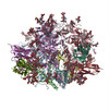 5fuuMC 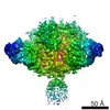 3309C 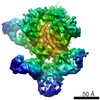 3312C M: atomic model generated by this map C: citing same article ( |
|---|---|
| Similar structure data |

-Links
| EMDB pages | EMDB (EBI/PDBe) / EMDataResource |
|---|---|
| Related items in Molecule of the Month |


-Map
| File | Download / File: emd_3308.map.gz / Format: CCP4 / Size: 62.5 MB / Type: IMAGE STORED AS FLOATING POINT NUMBER (4 BYTES) | ||||||||||||||||||||||||||||||||||||||||||||||||||||||||||||||||||||
|---|---|---|---|---|---|---|---|---|---|---|---|---|---|---|---|---|---|---|---|---|---|---|---|---|---|---|---|---|---|---|---|---|---|---|---|---|---|---|---|---|---|---|---|---|---|---|---|---|---|---|---|---|---|---|---|---|---|---|---|---|---|---|---|---|---|---|---|---|---|
| Annotation | Wild type JR-FL EnvdCT in complex with PGT151, with Fab constant region and detergent micelle masked out. | ||||||||||||||||||||||||||||||||||||||||||||||||||||||||||||||||||||
| Projections & slices | Image control
Images are generated by Spider. | ||||||||||||||||||||||||||||||||||||||||||||||||||||||||||||||||||||
| Voxel size | X=Y=Z: 1.31 Å | ||||||||||||||||||||||||||||||||||||||||||||||||||||||||||||||||||||
| Density |
| ||||||||||||||||||||||||||||||||||||||||||||||||||||||||||||||||||||
| Symmetry | Space group: 1 | ||||||||||||||||||||||||||||||||||||||||||||||||||||||||||||||||||||
| Details | EMDB XML:
CCP4 map header:
| ||||||||||||||||||||||||||||||||||||||||||||||||||||||||||||||||||||
 Z (Sec.)
 Y (Row.)
 X (Col.)


-Supplemental data
- Sample components
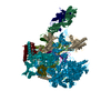
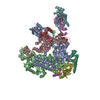
-Entire : Cleaved JR-FL EnvdCT in complex with PGT151 Fab
| Entire | Name: Cleaved JR-FL EnvdCT in complex with PGT151 Fab |
|---|---|
| Components |
|
-Supramolecule #1000: Cleaved JR-FL EnvdCT in complex with PGT151 Fab
| Supramolecule | Name: Cleaved JR-FL EnvdCT in complex with PGT151 Fab / type: sample / ID: 1000 / Oligomeric state: Two PGT151 Fabs bind one Env trimer / Number unique components: 2 |
|---|---|
| Molecular weight | Theoretical: 535 KDa |
-Macromolecule #1: HIV-1 Envelope glycoprotein
| Macromolecule | Name: HIV-1 Envelope glycoprotein / type: protein_or_peptide / ID: 1 / Name.synonym: HIV-1 Env Details: wild-type JR-FL Env trimer with the cytoplasmic tail truncated Number of copies: 1 / Oligomeric state: Trimer / Recombinant expression: Yes |
|---|---|
| Source (natural) | Organism: Human Immunodeficiency Virus-1 / Strain: JR-FL / synonym: HIV-1 |
| Molecular weight | Theoretical: 435 KDa |
| Recombinant expression | Organism: Mammalian (mammals) / Recombinant strain: Human / Recombinant cell: HEK293F / Recombinant plasmid: phCMV3 |
-Macromolecule #2: Immunoglobulin G PGT151
| Macromolecule | Name: Immunoglobulin G PGT151 / type: protein_or_peptide / ID: 2 / Name.synonym: IgG PGT151 / Details: PGT151 cleaved into Fab / Number of copies: 2 / Oligomeric state: Heterodimer / Recombinant expression: Yes |
|---|---|
| Source (natural) | Organism: Homo sapiens (human) / synonym: Human |
| Molecular weight | Theoretical: 50 KDa |
| Recombinant expression | Organism: Mammalian (mammals) / Recombinant strain: Human / Recombinant cell: HEK293F |
-Experimental details
-Structure determination
| Method | cryo EM |
|---|---|
 Processing Processing | single particle reconstruction |
| Aggregation state | particle |
-Sample preparation
| Concentration | 3.5 mg/mL |
|---|---|
| Buffer | pH: 7.4 Details: 50 mM Tris pH 7.4, 150 mM NaCl, 0.1% DDM, 0.03 mg/mL sodium deoxycholate |
| Grid | Details: 400 mesh C-Flat, CF-2/2-4C, plasma cleaned for 5 seconds |
| Vitrification | Cryogen name: ETHANE / Instrument: HOMEMADE PLUNGER Details: Samples were treated with biobeads prior to freezing Method: Grids were manually plunged at RT. |
- Electron microscopy #1
| Microscopy ID | 1 |
|---|---|
| Microscope | FEI TITAN KRIOS |
| Alignment procedure | Legacy - Astigmatism: Objective astigmatism corrected at 22,500x magnification. |
| Date | Nov 8, 2014 |
| Image recording | Category: CCD / Film or detector model: GATAN K2 SUMMIT (4k x 4k) / Digitization - Sampling interval: 5.0 µm / Number real images: 8759 / Average electron dose: 35.1 e/Å2 Details: Each full dose image is an aligned stack of frames recorded each using a dose of ~10 e-/Angstrom^2/sec. |
| Tilt angle min | 0 |
| Electron beam | Acceleration voltage: 300 kV / Electron source:  FIELD EMISSION GUN FIELD EMISSION GUN |
| Electron optics | Calibrated magnification: 22500 / Illumination mode: FLOOD BEAM / Imaging mode: BRIGHT FIELD / Cs: 2.7 mm / Nominal defocus max: 2.5 µm / Nominal defocus min: 1.5 µm / Nominal magnification: 22500 |
| Sample stage | Specimen holder model: FEI TITAN KRIOS AUTOGRID HOLDER |
| Experimental equipment |  Model: Titan Krios / Image courtesy: FEI Company |
-Electron microscopy #2
| Microscopy ID | 2 |
|---|---|
| Microscope | FEI TITAN KRIOS |
| Alignment procedure | Legacy - Astigmatism: Objective astigmatism corrected at 22,500x magnification. |
| Date | Oct 24, 2014 |
| Image recording | Category: CCD / Film or detector model: GATAN K2 SUMMIT (4k x 4k) / Digitization - Sampling interval: 5.0 µm / Number real images: 8759 / Average electron dose: 33.7 e/Å2 Details: Each full dose image is an aligned stack of frames recorded each using a dose of ~10 e-/Angstrom^2/sec. |
| Tilt angle min | 0 |
| Electron beam | Acceleration voltage: 300 kV / Electron source: FIELD EMISSION GUN |
| Electron optics | Calibrated magnification: 22500 / Illumination mode: FLOOD BEAM / Imaging mode: BRIGHT FIELD / Cs: 2.7 mm / Nominal defocus max: 3.5 µm / Nominal defocus min: 1.2 µm / Nominal magnification: 22500 |
| Sample stage | Specimen holder model: FEI TITAN KRIOS AUTOGRID HOLDER |
| Experimental equipment | Model: Titan Krios / Image courtesy: FEI Company |
-Electron microscopy #3
| Microscopy ID | 3 |
|---|---|
| Microscope | FEI TITAN KRIOS |
| Alignment procedure | Legacy - Astigmatism: Objective astigmatism corrected at 22,500x magnification. |
| Date | Sep 11, 2014 |
| Image recording | Category: CCD / Film or detector model: GATAN K2 SUMMIT (4k x 4k) / Digitization - Sampling interval: 5.0 µm / Number real images: 8759 / Average electron dose: 32.2 e/Å2 Details: Each full dose image is an aligned stack of frames recorded each using a dose of ~10 e-/Angstrom^2/sec. |
| Tilt angle min | 0 |
| Electron beam | Acceleration voltage: 300 kV / Electron source: FIELD EMISSION GUN |
| Electron optics | Calibrated magnification: 22500 / Illumination mode: FLOOD BEAM / Imaging mode: BRIGHT FIELD / Cs: 2.7 mm / Nominal defocus max: 4.0 µm / Nominal defocus min: 1.5 µm / Nominal magnification: 22500 |
| Sample stage | Specimen holder model: FEI TITAN KRIOS AUTOGRID HOLDER |
| Experimental equipment | Model: Titan Krios / Image courtesy: FEI Company |
-Electron microscopy #4
| Microscopy ID | 4 |
|---|---|
| Microscope | FEI TITAN KRIOS |
| Alignment procedure | Legacy - Astigmatism: Objective astigmatism corrected at 22,500x magnification. |
| Date | Feb 27, 2015 |
| Image recording | Category: CCD / Film or detector model: GATAN K2 SUMMIT (4k x 4k) / Digitization - Sampling interval: 5.0 µm / Number real images: 8759 / Average electron dose: 32.4 e/Å2 Details: Each full dose image is an aligned stack of frames recorded each using a dose of ~10 e-/Angstrom^2/sec. |
| Tilt angle min | 0 |
| Electron beam | Acceleration voltage: 300 kV / Electron source: FIELD EMISSION GUN |
| Electron optics | Calibrated magnification: 22500 / Illumination mode: FLOOD BEAM / Imaging mode: BRIGHT FIELD / Cs: 2.7 mm / Nominal defocus max: 3.0 µm / Nominal defocus min: 1.2 µm / Nominal magnification: 22500 |
| Sample stage | Specimen holder model: FEI TITAN KRIOS AUTOGRID HOLDER |
| Experimental equipment | Model: Titan Krios / Image courtesy: FEI Company |
-Image processing
| Details | At the end of refinement which gave the 4.3 Angstrom resolution structure, a mask was applied to exclude the flexible micelle and Fab constant regions, following which the model refined for an additional 3 iterations (until convergence). |
|---|---|
| CTF correction | Details: Each micrograph |
| Final reconstruction | Applied symmetry - Point group: C1 (asymmetric) / Algorithm: OTHER / Resolution.type: BY AUTHOR / Resolution: 4.19 Å / Resolution method: OTHER / Software - Name: Relion / Number images used: 201386 |
-Atomic model buiding 1
| Initial model | PDB ID: Chain - #0 - Chain ID: B / Chain - #1 - Chain ID: G |
|---|---|
| Software | Name: Chimera |
| Details | The trimer was used as an initial model for model building and refinement. |
| Refinement | Space: REAL / Protocol: RIGID BODY FIT |
| Output model | PDB-5fuu: |
-Atomic model buiding 2
| Initial model | PDB ID: Chain - #0 - Chain ID: H / Chain - #1 - Chain ID: L |
|---|---|
| Software | Name: Chimera |
| Details | The Fab was used as an initial model for model building and refinement. |
| Refinement | Space: REAL / Protocol: RIGID BODY FIT |
| Output model | PDB-5fuu: |