[English] 日本語
 Yorodumi
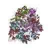
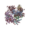
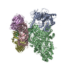
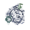
- PDB-5fuu: Ectodomain of cleaved wild type JR-FL EnvdCT trimer in complex wi... -
+ Open data
- Basic information
| Entry | Database: PDB / ID: 5fuu | |||||||||
|---|---|---|---|---|---|---|---|---|---|---|
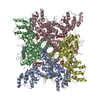
| Title | Ectodomain of cleaved wild type JR-FL EnvdCT trimer in complex with PGT151 Fab | |||||||||
 Components Components |
| |||||||||
 Keywords Keywords | VIRAL PROTEIN / HIV-1 / ENV / PGT151 / BROADLY NEUTRALIZING ANTIBODY | |||||||||
| Function / homology |  Function and homology information Function and homology informationsymbiont-mediated perturbation of host defense response / positive regulation of plasma membrane raft polarization / positive regulation of receptor clustering / host cell endosome membrane / clathrin-dependent endocytosis of virus by host cell / viral protein processing / fusion of virus membrane with host plasma membrane / fusion of virus membrane with host endosome membrane / viral envelope / virion attachment to host cell ...symbiont-mediated perturbation of host defense response / positive regulation of plasma membrane raft polarization / positive regulation of receptor clustering / host cell endosome membrane / clathrin-dependent endocytosis of virus by host cell / viral protein processing / fusion of virus membrane with host plasma membrane / fusion of virus membrane with host endosome membrane / viral envelope / virion attachment to host cell / host cell plasma membrane / virion membrane / structural molecule activity / identical protein binding / membrane Similarity search - Function | |||||||||
| Biological species |   HUMAN IMMUNODEFICIENCY VIRUS 1 HUMAN IMMUNODEFICIENCY VIRUS 1 HOMO SAPIENS (human) HOMO SAPIENS (human) | |||||||||
| Method | ELECTRON MICROSCOPY / single particle reconstruction / cryo EM / Resolution: 4.19 Å | |||||||||
 Authors Authors | Lee, J.H. / Ward, A.B. | |||||||||
 Citation Citation |  Journal: Science / Year: 2016 Journal: Science / Year: 2016Title: Cryo-EM structure of a native, fully glycosylated, cleaved HIV-1 envelope trimer. Authors: Jeong Hyun Lee / Gabriel Ozorowski / Andrew B Ward /  Abstract: The envelope glycoprotein trimer (Env) on the surface of HIV-1 recognizes CD4(+) T cells and mediates viral entry. During this process, Env undergoes substantial conformational rearrangements, making ...The envelope glycoprotein trimer (Env) on the surface of HIV-1 recognizes CD4(+) T cells and mediates viral entry. During this process, Env undergoes substantial conformational rearrangements, making it difficult to study in its native state. Soluble stabilized trimers have provided valuable insights into the Env structure, but they lack the hydrophobic membrane proximal external region (MPER, an important target of broadly neutralizing antibodies), the transmembrane domain, and the cytoplasmic tail. Here we present (i) a cryogenic electron microscopy (cryo-EM) structure of a clade B virus Env, which lacks only the cytoplasmic tail and is stabilized by the broadly neutralizing antibody PGT151, at a resolution of 4.2 angstroms and (ii) a reconstruction of this form of Env in complex with PGT151 and MPER-targeting antibody 10E8 at a resolution of 8.8 angstroms. These structures provide new insights into the wild-type Env structure. | |||||||||
| History |
|
- Structure visualization
| Movie |
 Movie viewer Movie viewer |
|---|---|
| Structure viewer | Molecule:  Molmil Molmil Jmol/JSmol Jmol/JSmol |
- Downloads & links
- Download
| PDBx/mmCIF format |  5fuu.cif.gz 5fuu.cif.gz | 466.3 KB | Display |  PDBx/mmCIF format PDBx/mmCIF format |
|---|---|---|---|---|
| PDB format |  pdb5fuu.ent.gz pdb5fuu.ent.gz | Display |  PDB format PDB format | |
| PDBx/mmJSON format |  5fuu.json.gz 5fuu.json.gz | Tree view |  PDBx/mmJSON format PDBx/mmJSON format | |
| Others |  Other downloads Other downloads |
-Validation report
| Summary document |  5fuu_validation.pdf.gz 5fuu_validation.pdf.gz | 4.4 MB | Display |  wwPDB validaton report wwPDB validaton report |
|---|---|---|---|---|
| Full document |  5fuu_full_validation.pdf.gz 5fuu_full_validation.pdf.gz | 4.5 MB | Display | |
| Data in XML |  5fuu_validation.xml.gz 5fuu_validation.xml.gz | 69.5 KB | Display | |
| Data in CIF |  5fuu_validation.cif.gz 5fuu_validation.cif.gz | 106.2 KB | Display | |
| Arichive directory |  https://data.pdbj.org/pub/pdb/validation_reports/fu/5fuu https://data.pdbj.org/pub/pdb/validation_reports/fu/5fuu ftp://data.pdbj.org/pub/pdb/validation_reports/fu/5fuu ftp://data.pdbj.org/pub/pdb/validation_reports/fu/5fuu | HTTPS FTP |
-Related structure data
| Related structure data |  3308MC  3309C  3312C M: map data used to model this data C: citing same article ( |
|---|---|
| Similar structure data |
- Links
Links
- Assembly
| Deposited unit | 
|
|---|---|
| 1 |
|
- Components
-HIV-1 ENVELOPE GLYCOPROTEIN ... , 2 types, 6 molecules ACEBDF
| #1: Protein | Mass: 53342.355 Da / Num. of mol.: 3 / Fragment: GP120, RESIDUES 30-502 / Mutation: YES Source method: isolated from a genetically manipulated source Details: GP120 OF JR-FL ENV / Source: (gene. exp.)   HUMAN IMMUNODEFICIENCY VIRUS 1 / Gene: ENV / Plasmid: PHCMV3 / Cell line (production host): HEK293 / Production host: HUMAN IMMUNODEFICIENCY VIRUS 1 / Gene: ENV / Plasmid: PHCMV3 / Cell line (production host): HEK293 / Production host:  HOMO SAPIENS (human) / References: UniProt: Q75760, UniProt: Q6BC19*PLUS HOMO SAPIENS (human) / References: UniProt: Q75760, UniProt: Q6BC19*PLUS#2: Protein | Mass: 17243.592 Da / Num. of mol.: 3 / Fragment: GP41, RESIDUES 503-655 Source method: isolated from a genetically manipulated source Details: GP41 ECTODOMAIN OF JR-FL ENV / Source: (gene. exp.)   HUMAN IMMUNODEFICIENCY VIRUS 1 / Gene: ENV / Plasmid: PHCMV3 / Cell line (production host): HEK293 / Production host: HUMAN IMMUNODEFICIENCY VIRUS 1 / Gene: ENV / Plasmid: PHCMV3 / Cell line (production host): HEK293 / Production host:  HOMO SAPIENS (human) / References: UniProt: Q6BC19 HOMO SAPIENS (human) / References: UniProt: Q6BC19 |
|---|
-Antibody , 2 types, 4 molecules HMLN
| #3: Antibody | Mass: 26087.438 Da / Num. of mol.: 2 / Fragment: FAB HEAVY CHAIN VARIABLE REGION, RESIDUES 1-218 Source method: isolated from a genetically manipulated source Details: PGT151 CLEAVED INTO FAB / Source: (gene. exp.)  HOMO SAPIENS (human) / Plasmid: PHCMV3 / Cell line (production host): HEK293 / Production host: HOMO SAPIENS (human) / Plasmid: PHCMV3 / Cell line (production host): HEK293 / Production host:  HOMO SAPIENS (human) HOMO SAPIENS (human)#4: Antibody | Mass: 24057.809 Da / Num. of mol.: 2 / Fragment: FAB LIGHT CHAIN VARIABLE REGION, RESIDUES 1-214 Source method: isolated from a genetically manipulated source Details: PGT151 CLEAVED INTO FAB / Source: (gene. exp.)  HOMO SAPIENS (human) / Plasmid: PHCMV3 / Cell line (production host): HEK293 / Production host: HOMO SAPIENS (human) / Plasmid: PHCMV3 / Cell line (production host): HEK293 / Production host:  HOMO SAPIENS (human) HOMO SAPIENS (human) |
|---|
-Sugars , 12 types, 64 molecules 
| #5: Polysaccharide | 2-acetamido-2-deoxy-beta-D-glucopyranose-(1-4)-2-acetamido-2-deoxy-beta-D-glucopyranose Source method: isolated from a genetically manipulated source #6: Polysaccharide | beta-D-mannopyranose-(1-4)-2-acetamido-2-deoxy-beta-D-glucopyranose-(1-4)-2-acetamido-2-deoxy-beta- ...beta-D-mannopyranose-(1-4)-2-acetamido-2-deoxy-beta-D-glucopyranose-(1-4)-2-acetamido-2-deoxy-beta-D-glucopyranose Source method: isolated from a genetically manipulated source #7: Polysaccharide | Source method: isolated from a genetically manipulated source #8: Polysaccharide | Source method: isolated from a genetically manipulated source #9: Polysaccharide | beta-D-galactopyranose-(1-4)-2-acetamido-2-deoxy-beta-D-glucopyranose-(1-2)-[beta-D-galactopyranose- ...beta-D-galactopyranose-(1-4)-2-acetamido-2-deoxy-beta-D-glucopyranose-(1-2)-[beta-D-galactopyranose-(1-4)-2-acetamido-2-deoxy-beta-D-glucopyranose-(1-4)]alpha-D-mannopyranose-(1-3)-[beta-D-galactopyranose-(1-4)-2-acetamido-2-deoxy-beta-D-glucopyranose-(1-2)-[2-acetamido-2-deoxy-beta-D-glucopyranose-(1-6)]alpha-D-mannopyranose-(1-6)]beta-D-mannopyranose-(1-4)-2-acetamido-2-deoxy-beta-D-glucopyranose-(1-4)-[alpha-L-fucopyranose-(1-6)]2-acetamido-2-deoxy-beta-D-glucopyranose | Source method: isolated from a genetically manipulated source #10: Polysaccharide | alpha-D-mannopyranose-(1-3)-[alpha-D-mannopyranose-(1-6)]beta-D-mannopyranose-(1-4)-2-acetamido-2- ...alpha-D-mannopyranose-(1-3)-[alpha-D-mannopyranose-(1-6)]beta-D-mannopyranose-(1-4)-2-acetamido-2-deoxy-beta-D-glucopyranose-(1-4)-2-acetamido-2-deoxy-beta-D-glucopyranose | Source method: isolated from a genetically manipulated source #11: Polysaccharide | Source method: isolated from a genetically manipulated source #12: Polysaccharide | 2-acetamido-2-deoxy-beta-D-glucopyranose-(1-4)-[alpha-L-fucopyranose-(1-6)]2-acetamido-2-deoxy-beta- ...2-acetamido-2-deoxy-beta-D-glucopyranose-(1-4)-[alpha-L-fucopyranose-(1-6)]2-acetamido-2-deoxy-beta-D-glucopyranose | Source method: isolated from a genetically manipulated source #13: Polysaccharide | alpha-D-mannopyranose-(1-2)-alpha-D-mannopyranose-(1-3)-[alpha-D-mannopyranose-(1-6)]beta-D- ...alpha-D-mannopyranose-(1-2)-alpha-D-mannopyranose-(1-3)-[alpha-D-mannopyranose-(1-6)]beta-D-mannopyranose-(1-4)-2-acetamido-2-deoxy-beta-D-glucopyranose-(1-4)-2-acetamido-2-deoxy-beta-D-glucopyranose | Source method: isolated from a genetically manipulated source #14: Polysaccharide | alpha-D-mannopyranose-(1-6)-beta-D-mannopyranose-(1-4)-2-acetamido-2-deoxy-beta-D-glucopyranose-(1- ...alpha-D-mannopyranose-(1-6)-beta-D-mannopyranose-(1-4)-2-acetamido-2-deoxy-beta-D-glucopyranose-(1-4)-2-acetamido-2-deoxy-beta-D-glucopyranose | Source method: isolated from a genetically manipulated source #15: Polysaccharide | beta-D-galactopyranose-(1-4)-2-acetamido-2-deoxy-beta-D-glucopyranose-(1-2)-[beta-D-galactopyranose- ...beta-D-galactopyranose-(1-4)-2-acetamido-2-deoxy-beta-D-glucopyranose-(1-2)-[beta-D-galactopyranose-(1-4)-2-acetamido-2-deoxy-beta-D-glucopyranose-(1-4)]alpha-D-mannopyranose-(1-3)-[2-acetamido-2-deoxy-beta-D-glucopyranose-(1-2)-[2-acetamido-2-deoxy-beta-D-glucopyranose-(1-6)]alpha-D-mannopyranose-(1-6)]beta-D-mannopyranose-(1-4)-2-acetamido-2-deoxy-beta-D-glucopyranose-(1-4)-[alpha-L-fucopyranose-(1-6)]2-acetamido-2-deoxy-beta-D-glucopyranose | Source method: isolated from a genetically manipulated source #16: Sugar | ChemComp-NAG / |
|---|
-Details
| Has protein modification | Y |
|---|---|
| Sequence details | V31 WAS FOUND TO HAVE MUTATED TO T31 WHEN THE PLASMID WAS SEQUENCED |
-Experimental details
-Experiment
| Experiment | Method: ELECTRON MICROSCOPY |
|---|---|
| EM experiment | Aggregation state: PARTICLE / 3D reconstruction method: single particle reconstruction |
- Sample preparation
| Component | Name: JR-FL ENVDCT IN COMPLEX WITH PGT151 FAB / Type: COMPLEX |
|---|---|
| Buffer solution | Name: 50 MM TRIS, 150 MM NACL, 0.1% DDM, 0.03 MG/ML SODIUM DEOXYCHOLATE pH: 7.4 Details: 50 MM TRIS, 150 MM NACL, 0.1% DDM, 0.03 MG/ML SODIUM DEOXYCHOLATE |
| Specimen | Conc.: 3.5 mg/ml / Embedding applied: NO / Shadowing applied: NO / Staining applied: NO / Vitrification applied: YES |
| Specimen support | Details: HOLEY CARBON |
| Vitrification | Instrument: HOMEMADE PLUNGER / Cryogen name: ETHANE Details: SAMPLES WERE PLUNGED INTO LIQUID ETHANE USING A MANUAL PLUNGER AT RT. |
- Electron microscopy imaging
| Experimental equipment |  Model: Titan Krios / Image courtesy: FEI Company |
|---|---|
| Microscopy | Model: FEI TITAN KRIOS / Date: Feb 27, 2015 |
| Electron gun | Electron source:  FIELD EMISSION GUN / Accelerating voltage: 300 kV / Illumination mode: FLOOD BEAM FIELD EMISSION GUN / Accelerating voltage: 300 kV / Illumination mode: FLOOD BEAM |
| Electron lens | Mode: BRIGHT FIELD / Nominal magnification: 22500 X / Calibrated magnification: 22500 X / Nominal defocus max: 3000 nm / Nominal defocus min: 1200 nm / Cs: 2.7 mm |
| Specimen holder | Tilt angle min: 0 ° |
| Image recording | Electron dose: 32.4 e/Å2 / Film or detector model: GATAN K2 SUMMIT (4k x 4k) |
- Processing
| EM software |
| ||||||||||||
|---|---|---|---|---|---|---|---|---|---|---|---|---|---|
| CTF correction | Details: WHOLE MICROGRAPH | ||||||||||||
| Symmetry | Point symmetry: C1 (asymmetric) | ||||||||||||
| 3D reconstruction | Resolution: 4.19 Å / Num. of particles: 201386 / Nominal pixel size: 1.31 Å / Actual pixel size: 1.31 Å Details: SUBMISSION BASED ON EXPERIMENTAL DATA FROM EMDB EMD-3308. (DEPOSITION ID: 14201). Symmetry type: POINT | ||||||||||||
| Atomic model building | Protocol: OTHER / Space: REAL / Details: REFINEMENT PROTOCOL--EM | ||||||||||||
| Refinement | Highest resolution: 4.19 Å | ||||||||||||
| Refinement step | Cycle: LAST / Highest resolution: 4.2 Å
|
 Movie
Movie Controller





 PDBj
PDBj


