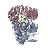
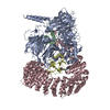
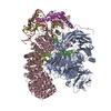
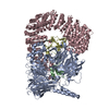
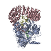
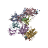
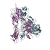
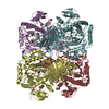
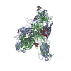
ジャーナル: Nat Commun / 年: 2021 タイトル: Structural basis of intron selection by U2 snRNP in the presence of covalent inhibitors. 著者: Constantin Cretu / Patricia Gee / Xiang Liu / Anant Agrawal / Tuong-Vi Nguyen / Arun K Ghosh / Andrew Cook / Melissa Jurica / Nicholas A Larsen / Vladimir Pena / 要旨: Intron selection during the formation of prespliceosomes is a critical event in pre-mRNA splicing. Chemical modulation of intron selection has emerged as a route for cancer therapy. Splicing ...Intron selection during the formation of prespliceosomes is a critical event in pre-mRNA splicing. Chemical modulation of intron selection has emerged as a route for cancer therapy. Splicing modulators alter the splicing patterns in cells by binding to the U2 snRNP (small nuclear ribonucleoprotein)-a complex chaperoning the selection of branch and 3' splice sites. Here we report crystal structures of the SF3B module of the U2 snRNP in complex with spliceostatin and sudemycin FR901464 analogs, and the cryo-electron microscopy structure of a cross-exon prespliceosome-like complex arrested with spliceostatin A. The structures reveal how modulators inactivate the branch site in a sequence-dependent manner and stall an E-to-A prespliceosome intermediate by covalent coupling to a nucleophilic zinc finger belonging to the SF3B subunit PHF5A. These findings support a mechanism of intron recognition by the U2 snRNP as a toehold-mediated strand invasion and advance an unanticipated drug targeting concept.
 ムービー
ムービー コントローラー


 データを開く
 基本情報
基本情報 要素
要素 キーワード
キーワード 機能・相同性情報
 Homo sapiens (ヒト)
 X線回折 /
  データ登録者
 ドイツ, 1件
  引用
引用

 構造の表示
構造の表示 ダウンロードとリンク
 その他のダウンロード
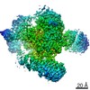


 PDBj
PDBj

 集合体
集合体
 Trichoplusia ni (イラクサキンウワバ) / 参照: UniProt: Q15393
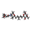


 分子量: 523.659 Da / 分子数: 1 / 由来タイプ: 合成 / 式: C28H45NO8 / タイプ: SUBJECT OF INVESTIGATION
 分子量: 65.409 Da / 分子数: 3 / 由来タイプ: 合成 / 式: Zn
 試料調製
試料調製 / ビームライン: X10SA / 波長: 1 Å
 解析
解析