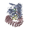
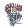
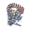
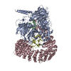
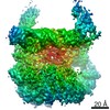
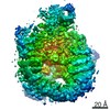
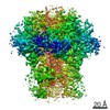
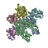
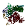
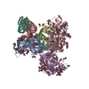
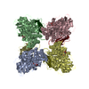
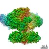
Journal: Nat Commun / Year: 2021 Title: Structural basis of intron selection by U2 snRNP in the presence of covalent inhibitors. Authors: Constantin Cretu / Patricia Gee / Xiang Liu / Anant Agrawal / Tuong-Vi Nguyen / Arun K Ghosh / Andrew Cook / Melissa Jurica / Nicholas A Larsen / Vladimir Pena / Abstract: Intron selection during the formation of prespliceosomes is a critical event in pre-mRNA splicing. Chemical modulation of intron selection has emerged as a route for cancer therapy. Splicing ...Intron selection during the formation of prespliceosomes is a critical event in pre-mRNA splicing. Chemical modulation of intron selection has emerged as a route for cancer therapy. Splicing modulators alter the splicing patterns in cells by binding to the U2 snRNP (small nuclear ribonucleoprotein)-a complex chaperoning the selection of branch and 3' splice sites. Here we report crystal structures of the SF3B module of the U2 snRNP in complex with spliceostatin and sudemycin FR901464 analogs, and the cryo-electron microscopy structure of a cross-exon prespliceosome-like complex arrested with spliceostatin A. The structures reveal how modulators inactivate the branch site in a sequence-dependent manner and stall an E-to-A prespliceosome intermediate by covalent coupling to a nucleophilic zinc finger belonging to the SF3B subunit PHF5A. These findings support a mechanism of intron recognition by the U2 snRNP as a toehold-mediated strand invasion and advance an unanticipated drug targeting concept.
History
Deposition
May 25, 2021
-
Header (metadata) release
Aug 4, 2021
-
Map release
Aug 4, 2021
-
Update
Nov 20, 2024
-
Current status
Nov 20, 2024
Processing site: PDBe / Status: Released
-
Structure visualization
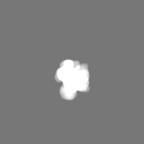
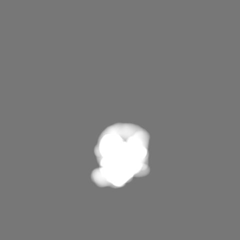
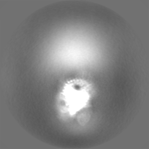
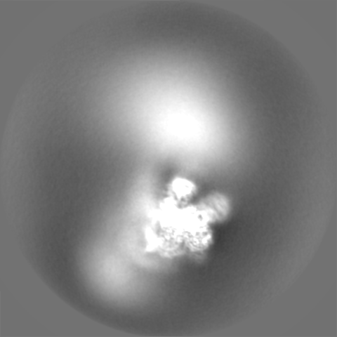
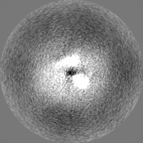
Movie
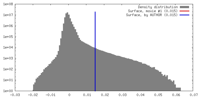
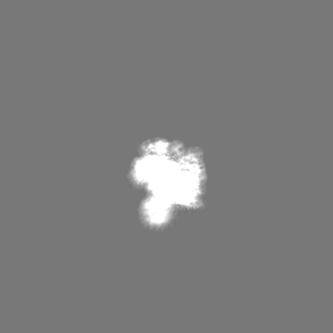
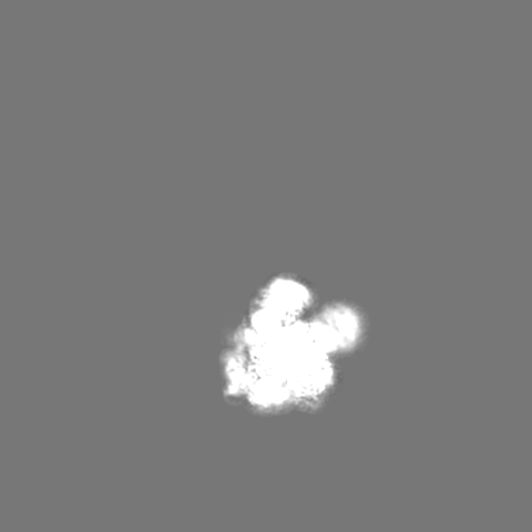
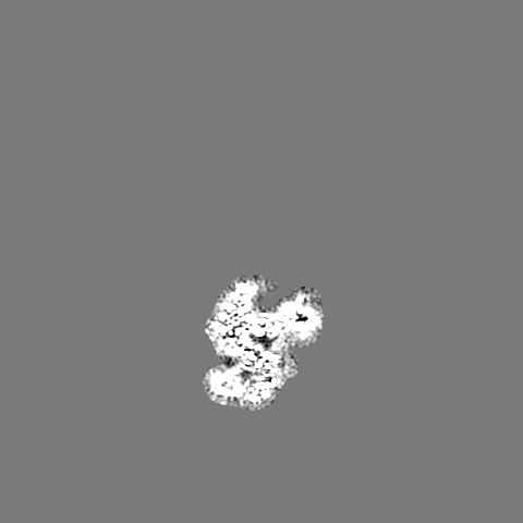
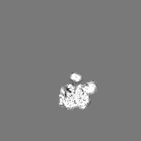
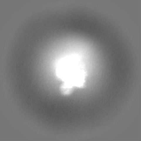
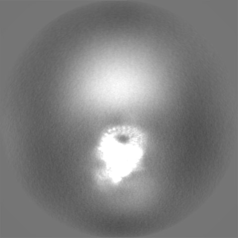
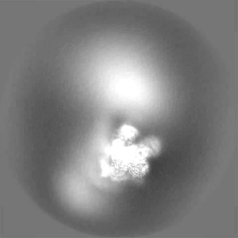
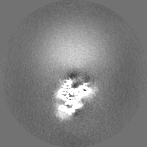
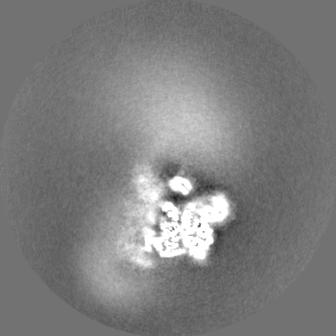
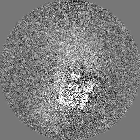
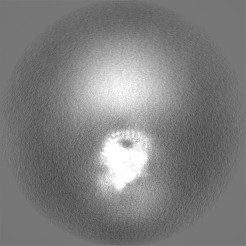
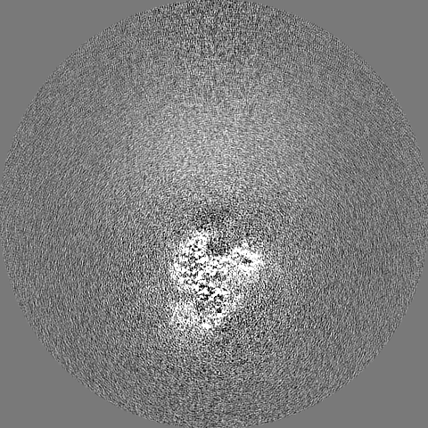
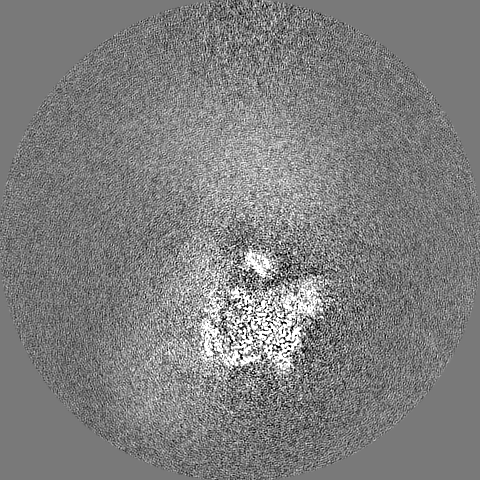
Surface view with section colored by density value
Cryogen name: ETHANE / Chamber humidity: 100 % / Chamber temperature: 277.15 K / Instrument: FEI VITROBOT MARK IV Details: The A3'-SSA complex was frozen in vitreous ice by applying ~2.2 uL crosslinked sample to both sides of a glow-discharged UltrAufoil R1.2/1.3 gold grid. The excess sample was blotted away for ...Details: The A3'-SSA complex was frozen in vitreous ice by applying ~2.2 uL crosslinked sample to both sides of a glow-discharged UltrAufoil R1.2/1.3 gold grid. The excess sample was blotted away for 2s using a blot force of 7 and the grid was snap frozen in liquid ethane cooled by liquid nitrogen..
-
Electron microscopy
Microscope
FEI TITAN KRIOS
Specialist optics
Energy filter - Name: GIF Quantum LS / Energy filter - Slit width: 20 eV
Image recording
Film or detector model: GATAN K3 (6k x 4k) / Number grids imaged: 1 / Number real images: 10494 / Average exposure time: 2.0 sec. / Average electron dose: 41.41 e/Å2
Electron beam
Acceleration voltage: 300 kV / Electron source: FIELD EMISSION GUN
In the structure databanks used in Yorodumi, some data are registered as the other names, "COVID-19 virus" and "2019-nCoV". Here are the details of the virus and the list of structure data.
Jan 31, 2019. EMDB accession codes are about to change! (news from PDBe EMDB page)
EMDB accession codes are about to change! (news from PDBe EMDB page)
The allocation of 4 digits for EMDB accession codes will soon come to an end. Whilst these codes will remain in use, new EMDB accession codes will include an additional digit and will expand incrementally as the available range of codes is exhausted. The current 4-digit format prefixed with “EMD-” (i.e. EMD-XXXX) will advance to a 5-digit format (i.e. EMD-XXXXX), and so on. It is currently estimated that the 4-digit codes will be depleted around Spring 2019, at which point the 5-digit format will come into force.
The EM Navigator/Yorodumi systems omit the EMD- prefix.
Related info.:Q: What is EMD? / ID/Accession-code notation in Yorodumi/EM Navigator
Yorodumi is a browser for structure data from EMDB, PDB, SASBDB, etc.
This page is also the successor to EM Navigator detail page, and also detail information page/front-end page for Omokage search.
The word "yorodu" (or yorozu) is an old Japanese word meaning "ten thousand". "mi" (miru) is to see.
Related info.:EMDB / PDB / SASBDB / Comparison of 3 databanks / Yorodumi Search / Aug 31, 2016. New EM Navigator & Yorodumi / Yorodumi Papers / Jmol/JSmol / Function and homology information / Changes in new EM Navigator and Yorodumi
 Movie
Movie Controller


 Open data
 Basic information
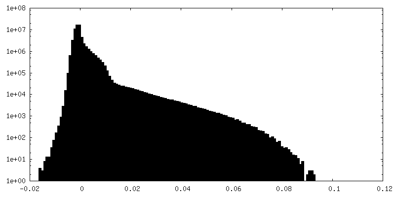
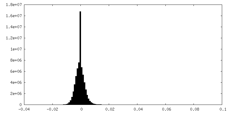
 Map data
 Sample
 Keywords
 Function and homology information
 Homo sapiens (human) /
  unidentified adenovirus
 Authors
 Germany, 1 items
  Citation

 Structure visualization
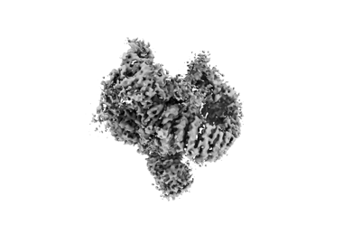
 Downloads & links
 emd_12994.png
 http://ftp.pdbj.org/pub/emdb/structures/EMD-12994


 Z (Sec.)
 Y (Row.)
 X (Col.)


 Sample components
 Processing
 Electron microscopy
 FIELD EMISSION GUN