 Movie
Movie Controller


+ Open data

- Basic information
| Entry | Database: PDB / ID: 1suv | ||||||
|---|---|---|---|---|---|---|---|
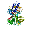
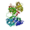
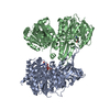
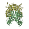
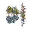
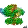
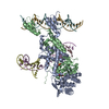
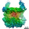
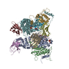
| Title | Structure of Human Transferrin Receptor-Transferrin Complex | ||||||
 Components Components |
| ||||||
 Keywords Keywords | METAL TRANSPORT / Protein Complex | ||||||
| Function / homology |  Function and homology information Function and homology informationtransferrin receptor activity / postsynaptic recycling endosome membrane / negative regulation of mitochondrial fusion / iron chaperone activity / positive regulation of isotype switching / transferrin receptor binding / Transferrin endocytosis and recycling / basal part of cell / response to manganese ion / Differentiation of Keratinocytes in Interfollicular Epidermis in Mammalian Skin ...transferrin receptor activity / postsynaptic recycling endosome membrane / negative regulation of mitochondrial fusion / iron chaperone activity / positive regulation of isotype switching / transferrin receptor binding / Transferrin endocytosis and recycling / basal part of cell / response to manganese ion / Differentiation of Keratinocytes in Interfollicular Epidermis in Mammalian Skin / response to iron ion / RND1 GTPase cycle / RND2 GTPase cycle / RHOB GTPase cycle / response to copper ion / RHOJ GTPase cycle / RHOC GTPase cycle / Golgi Associated Vesicle Biogenesis / RHOQ GTPase cycle / CDC42 GTPase cycle / RHOG GTPase cycle / RHOH GTPase cycle / RAC3 GTPase cycle / RHOA GTPase cycle / RAC2 GTPase cycle / response to retinoic acid / endocytic vesicle / regulation of postsynaptic membrane neurotransmitter receptor levels / transport across blood-brain barrier / positive regulation of B cell proliferation / RAC1 GTPase cycle / clathrin-coated pit / osteoclast differentiation / response to nutrient / positive regulation of T cell proliferation / ferric iron binding / receptor-mediated endocytosis / Hsp70 protein binding / basal plasma membrane / acute-phase response / Post-translational protein phosphorylation / iron ion transport / clathrin-coated endocytic vesicle membrane / regulation of protein stability / HFE-transferrin receptor complex / transferrin transport / receptor internalization / cellular response to iron ion / positive regulation of protein-containing complex assembly / ferrous iron binding / Iron uptake and transport / cellular response to xenobiotic stimulus / positive regulation of protein localization to nucleus / multicellular organismal-level iron ion homeostasis / positive regulation of receptor-mediated endocytosis / recycling endosome / Regulation of Insulin-like Growth Factor (IGF) transport and uptake by Insulin-like Growth Factor Binding Proteins (IGFBPs) / recycling endosome membrane / melanosome / Platelet degranulation / late endosome / Cargo recognition for clathrin-mediated endocytosis / positive regulation of proteasomal ubiquitin-dependent protein catabolic process / double-stranded RNA binding / antibacterial humoral response / Clathrin-mediated endocytosis / extracellular vesicle / virus receptor activity / cytoplasmic vesicle / secretory granule lumen / blood microparticle / vesicle / basolateral plasma membrane / intracellular iron ion homeostasis / transmembrane transporter binding / response to hypoxia / early endosome / positive regulation of canonical NF-kappaB signal transduction / cell surface receptor signaling pathway / lysosome / endosome / apical plasma membrane / endosome membrane / intracellular signal transduction / endoplasmic reticulum lumen / external side of plasma membrane / positive regulation of gene expression / protein kinase binding / negative regulation of apoptotic process / protein-containing complex binding / perinuclear region of cytoplasm / glutamatergic synapse / enzyme binding / cell surface / protein homodimerization activity / : / RNA binding / extracellular exosome / extracellular region / membrane Similarity search - Function | ||||||
| Biological species |  Homo sapiens (human) Homo sapiens (human) | ||||||
| Method | ELECTRON MICROSCOPY / single particle reconstruction / cryo EM / Resolution: 7.5 Å | ||||||
 Authors Authors | Cheng, Y. / Zak, O. / Aisen, P. / Harrison, S.C. / Walz, T. | ||||||
 Citation Citation | Journal: Cell / Year: 2004 Title: Structure of the human transferrin receptor-transferrin complex. Authors: Yifan Cheng / Olga Zak / Philip Aisen / Stephen C Harrison / Thomas Walz /  Abstract: Iron, insoluble as free Fe(3+) and toxic as free Fe(2+), is distributed through the body as Fe(3+) bound to transferrin (Tf) for delivery to cells by endocytosis of its complex with transferrin ...Iron, insoluble as free Fe(3+) and toxic as free Fe(2+), is distributed through the body as Fe(3+) bound to transferrin (Tf) for delivery to cells by endocytosis of its complex with transferrin receptor (TfR). Although much is understood of the transferrin endocytotic cycle, little has been uncovered of the molecular details underlying the formation of the receptor-transferrin complex. Using cryo-electron microscopy, we have produced a density map of the TfR-Tf complex at subnanometer resolution. An atomic model, obtained by fitting crystal structures of diferric Tf and the receptor ectodomain into the map, shows that the Tf N-lobe is sandwiched between the membrane and the TfR ectodomain and that the C-lobe abuts the receptor helical domain. When Tf binds receptor, its N-lobe moves by about 9 A with respect to its C-lobe. The structure of TfR-Tf complex helps account for known differences in the iron-release properties of free and receptor bound Tf. | ||||||
| History |
| ||||||
| Remark 999 | SEQUENCE NOT ALL THE CHAINS IN THE MODEL ARE HUMAN, ALTHOUGH THE PROTEINS USED TO DETERMINE THE 7.5 ...SEQUENCE NOT ALL THE CHAINS IN THE MODEL ARE HUMAN, ALTHOUGH THE PROTEINS USED TO DETERMINE THE 7.5 A STRUCTURE OF THE TFR-TF COMPLEX WERE ALL HUMAN. THE AUTHORS CREATED THE MODEL BY FITTING X-RAY CRYSTAL STRUCTURES INTO THEIR 7.5 A EM DENSITY MAP. SINCE THERE IS NO STRUCTURE FOR THE HUMAN TRANSFERRIN C-LOBE, THE AUTHORS OPTED TO USE THE C-LOBE FROM RABBIT TF (1JNF). THE OTHER TWO CHAINS ARE HUMAN (1CX8 - HUMAN TFR AND 1A8E - HUMAN TF N-LOBE). THE CHAINS E AND F MATCH SWS P19134, A RABBIT SOURCE. REGARDING THE CONFLICTS: BOTH SEQUENCE AND COORDINATES ARE FROM THE ORIGINAL PDB-FILES AND THE AUTHORS DID NOT MAKE ANY MODIFICATIONS TO IT. |
- Structure visualization
| Movie |
Movie viewer |
|---|---|
| Structure viewer | Molecule: MolmilJmol/JSmol |
- Downloads & links
-Download
| PDBx/mmCIF format | 1suv.cif.gz | 511.1 KB | Display | PDBx/mmCIF format |
|---|---|---|---|---|
| PDB format | pdb1suv.ent.gz | 411.9 KB | Display | PDB format |
| PDBx/mmJSON format | 1suv.json.gz | Tree view | PDBx/mmJSON format | |
| Others |  Other downloads Other downloads |
-Validation report
| Arichive directory | https://data.pdbj.org/pub/pdb/validation_reports/su/1suvftp://data.pdbj.org/pub/pdb/validation_reports/su/1suv | HTTPS FTP |
|---|
-Related structure data
| Related structure data | |
|---|---|
| Similar structure data |

-Links
 PDBj
PDBj


- Assembly
| Deposited unit | 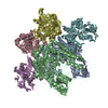
|
|---|---|
| 1 |
|
| Symmetry | Point symmetry: (Schoenflies symbol: C2 (2 fold cyclic)) |
-Components
| #1: Protein | Mass: 71622.961 Da / Num. of mol.: 2 Source method: isolated from a genetically manipulated source Source: (gene. exp.) Homo sapiens (human) / Gene: TFRC / Cell (production host): ovary / Production host:   Cricetulus griseus (Chinese hamster) / References: UniProt: P02786 Cricetulus griseus (Chinese hamster) / References: UniProt: P02786#2: Protein | Mass: 36408.414 Da / Num. of mol.: 2 / Fragment: repeat 1 Source method: isolated from a genetically manipulated source Source: (gene. exp.) Homo sapiens (human) / Gene: TF / Cell (production host): kidney / Production host: Cricetulus griseus (Chinese hamster) / References: UniProt: P02787#3: Protein | Mass: 38300.445 Da / Num. of mol.: 2 / Fragment: repeat 2 Source method: isolated from a genetically manipulated source Source: (gene. exp.) Homo sapiens (human) / Gene: TF / Cell (production host): kidney / Production host: Cricetulus griseus (Chinese hamster)#4: Chemical | ChemComp-CO3 /   Mass: 60.009 Da / Num. of mol.: 4 / Source method: obtained synthetically / Formula: CO3 Mass: 60.009 Da / Num. of mol.: 4 / Source method: obtained synthetically / Formula: CO3#5: Chemical | ChemComp-FE /   Mass: 55.845 Da / Num. of mol.: 4 / Source method: obtained synthetically / Formula: Fe Mass: 55.845 Da / Num. of mol.: 4 / Source method: obtained synthetically / Formula: FeHas protein modification | Y | |
|---|
-Experimental details
-Experiment
| Experiment | Method: ELECTRON MICROSCOPY |
|---|---|
| EM experiment | Aggregation state: PARTICLE / 3D reconstruction method: single particle reconstruction |
- Sample preparation
| Component | Name: Human Transferrin Receptor - Transferrin Complex / Type: COMPLEX |
|---|---|
| Buffer solution | pH: 7.4 |
| Specimen | Embedding applied: NO / Shadowing applied: NO / Staining applied: NO / Vitrification applied: YES |
- Electron microscopy imaging
| Experimental equipment |  Model: Tecnai F20 / Image courtesy: FEI Company |
|---|---|
| Microscopy | Model: FEI TECNAI F20 / Date: Oct 15, 2001 |
| Electron gun | Electron source:  FIELD EMISSION GUN / Accelerating voltage: 200 kV / Illumination mode: FLOOD BEAM FIELD EMISSION GUN / Accelerating voltage: 200 kV / Illumination mode: FLOOD BEAM |
| Electron lens | Mode: BRIGHT FIELD / Nominal magnification: 50000 X / Calibrated magnification: 51160 X / Nominal defocus max: 5000 nm / Nominal defocus min: 2500 nm / Cs: 2 mm |
| Specimen holder | Temperature: 93 K / Tilt angle max: 0 ° / Tilt angle min: 0 ° |
| Image recording | Electron dose: 20 e/Å2 / Film or detector model: KODAK SO-163 FILM |
- Processing
| CTF correction | Details: CTF correction for each particle | ||||||||||||||||||||||||||||
|---|---|---|---|---|---|---|---|---|---|---|---|---|---|---|---|---|---|---|---|---|---|---|---|---|---|---|---|---|---|
| Symmetry | Point symmetry: C2 (2 fold cyclic) | ||||||||||||||||||||||||||||
| 3D reconstruction | Method: Fourier Space reconstruction / Resolution: 7.5 Å / Nominal pixel size: 2.8 Å / Actual pixel size: 2.74 Å / Details: using program FREALIGN / Symmetry type: POINT | ||||||||||||||||||||||||||||
| Atomic model building | Protocol: RIGID BODY FIT / Space: REAL Target criteria: visual fit using program O followed by rigid body refinement using program MAVE Details: REFINEMENT PROTOCOL--rigid body | ||||||||||||||||||||||||||||
| Atomic model building |
| ||||||||||||||||||||||||||||
| Refinement step | Cycle: LAST
|
