[English] 日本語
 Yorodumi
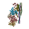
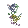
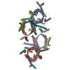
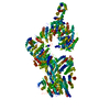
- PDB-6yg8: Cryo-EM structure of a BcsB pentamer in the context of an assembl... -
+ Open data
- Basic information
| Entry | Database: PDB / ID: 6yg8 | ||||||||||||||||||
|---|---|---|---|---|---|---|---|---|---|---|---|---|---|---|---|---|---|---|---|
| Title | Cryo-EM structure of a BcsB pentamer in the context of an assembled Bcs macrocomplex | ||||||||||||||||||
 Components Components | Bacterial cellulose secretion regulator BcsB | ||||||||||||||||||
 Keywords Keywords | SIGNALING PROTEIN / Bacterial biofilms / Bacterial cellulose / Bacterial secretion system / regulator protein | ||||||||||||||||||
| Function / homology | Cellulose synthase, subunit B / Cellulose synthase BcsB, bacterial / Bacterial cellulose synthase subunit / cellulose biosynthetic process / UDP-alpha-D-glucose metabolic process / plasma membrane / Cyclic di-GMP-binding protein / Cyclic di-GMP-binding protein Function and homology information Function and homology information | ||||||||||||||||||
| Biological species |  | ||||||||||||||||||
| Method | ELECTRON MICROSCOPY / single particle reconstruction / cryo EM / Resolution: 3 Å | ||||||||||||||||||
 Authors Authors | Zouhir, S. / Krasteva, P.V. | ||||||||||||||||||
| Funding support | 5items
| ||||||||||||||||||
 Citation Citation |  Journal: Sci Adv / Year: 2021 Journal: Sci Adv / Year: 2021Title: Architecture and regulation of an enterobacterial cellulose secretion system. Authors: Wiem Abidi / Samira Zouhir / Meryem Caleechurn / Stéphane Roche / Petya Violinova Krasteva /  Abstract: Many free-living and pathogenic enterobacteria secrete biofilm-promoting cellulose using a multicomponent, envelope-embedded Bcs secretion system under the control of intracellular second messenger c- ...Many free-living and pathogenic enterobacteria secrete biofilm-promoting cellulose using a multicomponent, envelope-embedded Bcs secretion system under the control of intracellular second messenger c-di-GMP. The molecular understanding of system assembly and cellulose secretion has been largely limited to the crystallographic studies of a distantly homologous BcsAB synthase tandem and a low-resolution reconstruction of an assembled macrocomplex that encompasses most of the inner membrane and cytosolic subunits and features an atypical layered architecture. Here, we present cryo-EM structures of the assembled Bcs macrocomplex, as well as multiple crystallographic snapshots of regulatory Bcs subcomplexes. The structural and functional data uncover the mechanism of asymmetric secretion system assembly and periplasmic crown polymerization and reveal unexpected subunit stoichiometry, multisite c-di-GMP recognition, and ATP-dependent regulation. | ||||||||||||||||||
| History |
|
- Structure visualization
| Movie |
 Movie viewer Movie viewer |
|---|---|
| Structure viewer | Molecule:  Molmil Molmil Jmol/JSmol Jmol/JSmol |
- Downloads & links
- Download
| PDBx/mmCIF format |  6yg8.cif.gz 6yg8.cif.gz | 532 KB | Display |  PDBx/mmCIF format PDBx/mmCIF format |
|---|---|---|---|---|
| PDB format |  pdb6yg8.ent.gz pdb6yg8.ent.gz | 440.3 KB | Display |  PDB format PDB format |
| PDBx/mmJSON format |  6yg8.json.gz 6yg8.json.gz | Tree view |  PDBx/mmJSON format PDBx/mmJSON format | |
| Others |  Other downloads Other downloads |
-Validation report
| Arichive directory |  https://data.pdbj.org/pub/pdb/validation_reports/yg/6yg8 https://data.pdbj.org/pub/pdb/validation_reports/yg/6yg8 ftp://data.pdbj.org/pub/pdb/validation_reports/yg/6yg8 ftp://data.pdbj.org/pub/pdb/validation_reports/yg/6yg8 | HTTPS FTP |
|---|
-Related structure data
| Related structure data |  10799MC 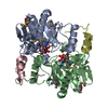 6yarC 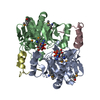 6yayC 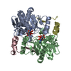 6yb3C 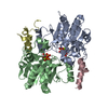 6yb5C 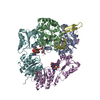 6ybbC 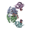 6ybuC C: citing same article ( M: map data used to model this data |
|---|---|
| Similar structure data |
- Links
Links
- Assembly
| Deposited unit | 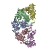
|
|---|---|
| 1 |
|
- Components
| #1: Protein | Mass: 86184.383 Da / Num. of mol.: 5 Source method: isolated from a genetically manipulated source Details: Bacterial cellulose synthesis subunit B / Source: (gene. exp.)  Gene: bcsB, A8C65_00280, AC789_1c39010, ACU57_05345, AM464_10380, BHS81_21120, BK292_24560, BON72_13325, BON75_10020, BON76_21165, BON94_21155, BZL31_14275, C5P01_24165, C9162_26260, CI641_010915, ...Gene: bcsB, A8C65_00280, AC789_1c39010, ACU57_05345, AM464_10380, BHS81_21120, BK292_24560, BON72_13325, BON75_10020, BON76_21165, BON94_21155, BZL31_14275, C5P01_24165, C9162_26260, CI641_010915, CWS33_18410, D3O91_11715, D9I18_15765, DQF57_09480, E0J34_09745, E0K84_22715, E2127_16410, E2128_18000, E2129_18135, E2134_17800, E5P22_13645, ELV08_08610, F1E03_18495, F1E19_10130, FRV13_18850 Production host:  Has protein modification | Y | |
|---|
-Experimental details
-Experiment
| Experiment | Method: ELECTRON MICROSCOPY |
|---|---|
| EM experiment | Aggregation state: PARTICLE / 3D reconstruction method: single particle reconstruction |
- Sample preparation
| Component |
| ||||||||||||||||||||||||||||
|---|---|---|---|---|---|---|---|---|---|---|---|---|---|---|---|---|---|---|---|---|---|---|---|---|---|---|---|---|---|
| Molecular weight | Value: 0.734 MDa / Experimental value: NO | ||||||||||||||||||||||||||||
| Source (natural) |
| ||||||||||||||||||||||||||||
| Source (recombinant) |
| ||||||||||||||||||||||||||||
| Buffer solution | pH: 8 | ||||||||||||||||||||||||||||
| Buffer component |
| ||||||||||||||||||||||||||||
| Specimen | Conc.: 0.8 mg/ml / Embedding applied: NO / Shadowing applied: NO / Staining applied: NO / Vitrification applied: YES | ||||||||||||||||||||||||||||
| Vitrification | Instrument: FEI VITROBOT MARK IV / Cryogen name: ETHANE / Humidity: 100 % / Chamber temperature: 277 K |
- Electron microscopy imaging
| Experimental equipment |  Model: Titan Krios / Image courtesy: FEI Company |
|---|---|
| Microscopy | Model: FEI TITAN KRIOS |
| Electron gun | Electron source:  FIELD EMISSION GUN / Accelerating voltage: 300 kV / Illumination mode: FLOOD BEAM FIELD EMISSION GUN / Accelerating voltage: 300 kV / Illumination mode: FLOOD BEAM |
| Electron lens | Mode: BRIGHT FIELD / Nominal magnification: 130000 X / Nominal defocus max: 2750 nm / Nominal defocus min: 750 nm / Cs: 2.7 mm / C2 aperture diameter: 100 µm |
| Image recording | Electron dose: 1.2 e/Å2 / Detector mode: COUNTING / Film or detector model: GATAN K2 SUMMIT (4k x 4k) |
| EM imaging optics | Energyfilter name: GIF Quantum LS |
- Processing
| Software | Name: PHENIX / Version: 1.16_3549: / Classification: refinement | |||||||||||||||||||||||||||||||||||||||||||||
|---|---|---|---|---|---|---|---|---|---|---|---|---|---|---|---|---|---|---|---|---|---|---|---|---|---|---|---|---|---|---|---|---|---|---|---|---|---|---|---|---|---|---|---|---|---|---|
| EM software |
| |||||||||||||||||||||||||||||||||||||||||||||
| Image processing | Details: A total of 9,129 movies recorded on the CM01 Titan Krios transmission electron microscope (Thermo Fisher Scientific) at the ESRF Grenoble operated at 300 kV and equipped with a Gatan K2 ...Details: A total of 9,129 movies recorded on the CM01 Titan Krios transmission electron microscope (Thermo Fisher Scientific) at the ESRF Grenoble operated at 300 kV and equipped with a Gatan K2 Summit direct electron detector and a GIF Quantum LS imaging filter | |||||||||||||||||||||||||||||||||||||||||||||
| CTF correction | Details: Gctf through the cryoSPARC v2 interface. / Type: PHASE FLIPPING AND AMPLITUDE CORRECTION | |||||||||||||||||||||||||||||||||||||||||||||
| Symmetry | Point symmetry: C1 (asymmetric) | |||||||||||||||||||||||||||||||||||||||||||||
| 3D reconstruction | Resolution: 3 Å / Resolution method: FSC 0.143 CUT-OFF / Num. of particles: 576455 / Num. of class averages: 1 / Symmetry type: POINT | |||||||||||||||||||||||||||||||||||||||||||||
| Atomic model building | Space: REAL | |||||||||||||||||||||||||||||||||||||||||||||
| Refine LS restraints |
|
 Movie
Movie Controller





 PDBj
PDBj
