 Movie
Movie Controller


+ Open data

- Basic information
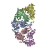
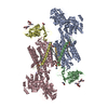
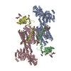
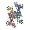
| Entry | Database: EMDB / ID: EMD-23146 | ||||||||||||
|---|---|---|---|---|---|---|---|---|---|---|---|---|---|
| Title | Bacterial cellulose synthase BcsB hexamer | ||||||||||||
 Map data Map data | |||||||||||||
 Sample Sample |
| ||||||||||||
 Keywords Keywords | bacterial cellulose / periplasmic / structural subunit / BIOSYNTHETIC PROTEIN | ||||||||||||
| Function / homology | Cellulose synthase, subunit B / Biofilm formation / Cellulose synthase BcsB, bacterial / Cellulose synthase BcsB, first CBM domain / cellulose biosynthetic process / UDP-alpha-D-glucose metabolic process / plasma membrane / Cyclic di-GMP-binding protein Function and homology information Function and homology information | ||||||||||||
| Biological species |  | ||||||||||||
| Method | single particle reconstruction / cryo EM / Resolution: 3.4 Å | ||||||||||||
 Authors Authors | Acheson JF / Zimmer J | ||||||||||||
| Funding support |  United States, 3 items United States, 3 items
| ||||||||||||
 Citation Citation | Journal: Nat Struct Mol Biol / Year: 2021 Title: Molecular organization of the E. coli cellulose synthase macrocomplex. Authors: Justin F Acheson / Ruoya Ho / Nicolette F Goularte / Lynette Cegelski / Jochen Zimmer / Abstract: Cellulose is frequently found in communities of sessile bacteria called biofilms. Escherichia coli and other enterobacteriaceae modify cellulose with phosphoethanolamine (pEtN) to promote host tissue ...Cellulose is frequently found in communities of sessile bacteria called biofilms. Escherichia coli and other enterobacteriaceae modify cellulose with phosphoethanolamine (pEtN) to promote host tissue adhesion. The E. coli pEtN cellulose biosynthesis machinery contains the catalytic BcsA-B complex that synthesizes and secretes cellulose, in addition to five other subunits. The membrane-anchored periplasmic BcsG subunit catalyzes pEtN modification. Here we present the structure of the roughly 1 MDa E. coli Bcs complex, consisting of one BcsA enzyme associated with six copies of BcsB, determined by single-particle cryo-electron microscopy. BcsB homo-oligomerizes primarily through interactions between its carbohydrate-binding domains as well as intermolecular beta-sheet formation. The BcsB hexamer creates a half spiral whose open side accommodates two BcsG subunits, directly adjacent to BcsA's periplasmic channel exit. The cytosolic BcsE and BcsQ subunits associate with BcsA's regulatory PilZ domain. The macrocomplex is a fascinating example of cellulose synthase specification. | ||||||||||||
| History |
|
- Structure visualization
| Movie |
Movie viewer |
|---|---|
| Structure viewer | EM map: SurfViewMolmilJmol/JSmol |
| Supplemental images |

- Downloads & links
-EMDB archive
| Map data | emd_23146.map.gz | 168 MB | EMDB map data format | |
|---|---|---|---|---|
| Header (meta data) | emd-23146-v30.xmlemd-23146.xml | 15.8 KB 15.8 KB | Display Display | EMDB header |
| FSC (resolution estimation) | emd_23146_fsc.xml | 12.5 KB | Display | FSC data file |
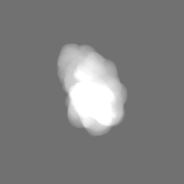
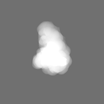
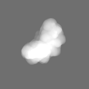
| Images | 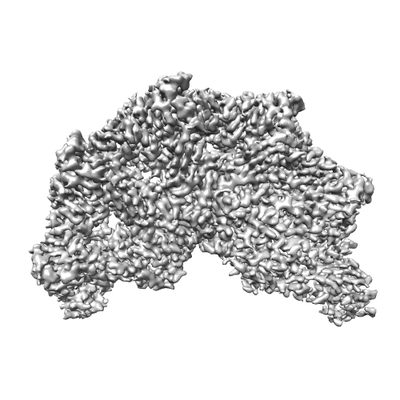 emd_23146.png emd_23146.png | 58.6 KB | ||
| Masks | emd_23146_msk_1.map | 178 MB | Mask map | |
| Filedesc metadata | emd-23146.cif.gz | 6.5 KB | ||
| Archive directory |  http://ftp.pdbj.org/pub/emdb/structures/EMD-23146ftp://ftp.pdbj.org/pub/emdb/structures/EMD-23146 http://ftp.pdbj.org/pub/emdb/structures/EMD-23146ftp://ftp.pdbj.org/pub/emdb/structures/EMD-23146 | HTTPS FTP |
-Related structure data
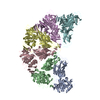
| Related structure data | 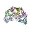 7l2zMC 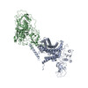 7lbyC M: atomic model generated by this map C: citing same article ( |
|---|---|
| Similar structure data | |
| EM raw data | EMPIAR-10626 (Title: Cryo EM Structure of the E. coli BcsB Hexamer / Data size: 837.2 Data #1: Unaligned multi-frame micrographs [micrographs - multiframe] Data #2: Unaligned movie frames [micrographs - multiframe] / Data #3: Unaligned movie frames [micrographs - multiframe] / Data #4: Unaligned movie frames [micrographs - multiframe]) EMPIAR-10627 (Title: Poly-alanine backbone model of E. coli BcsA bound to BcsBData size: 3.2 TB Data #1: Unaligned multi-frame micrographs [micrographs - multiframe] Data #2: Unaligned movie frames [micrographs - multiframe] / Data #3: Unaligned movie frames [micrographs - multiframe] / Data #4: Unaligned movie frames [micrographs - multiframe] / Data #5: Unaligned movie frames [micrographs - multiframe]) |


-Links
| EMDB pages | EMDB (EBI/PDBe) / EMDataResource |
|---|---|
| Related items in Molecule of the Month |

-Map
| File | Download / File: emd_23146.map.gz / Format: CCP4 / Size: 178 MB / Type: IMAGE STORED AS FLOATING POINT NUMBER (4 BYTES) | ||||||||||||||||||||||||||||||||||||||||||||||||||||||||||||
|---|---|---|---|---|---|---|---|---|---|---|---|---|---|---|---|---|---|---|---|---|---|---|---|---|---|---|---|---|---|---|---|---|---|---|---|---|---|---|---|---|---|---|---|---|---|---|---|---|---|---|---|---|---|---|---|---|---|---|---|---|---|
| Projections & slices | Image control
Images are generated by Spider. | ||||||||||||||||||||||||||||||||||||||||||||||||||||||||||||
| Voxel size | X=Y=Z: 1.08 Å | ||||||||||||||||||||||||||||||||||||||||||||||||||||||||||||
| Density |
| ||||||||||||||||||||||||||||||||||||||||||||||||||||||||||||
| Symmetry | Space group: 1 | ||||||||||||||||||||||||||||||||||||||||||||||||||||||||||||
| Details | EMDB XML:
CCP4 map header:
| ||||||||||||||||||||||||||||||||||||||||||||||||||||||||||||
 Z (Sec.)
 Y (Row.)
 X (Col.)


-Supplemental data
-Mask #1
| File | emd_23146_msk_1.map | ||||||||||||
|---|---|---|---|---|---|---|---|---|---|---|---|---|---|
| Projections & Slices |
| ||||||||||||
| Density Histograms |


- Sample components
-Entire : Bacterial cellulose synthase BcsB hexamer
| Entire | Name: Bacterial cellulose synthase BcsB hexamer |
|---|---|
| Components |
|
-Supramolecule #1: Bacterial cellulose synthase BcsB hexamer
| Supramolecule | Name: Bacterial cellulose synthase BcsB hexamer / type: complex / ID: 1 / Parent: 0 / Macromolecule list: all Details: Periplasmic domain of BcsB refined using masked local refinement and signal subtraction and from E coli BCS inner-membrane complex projections. |
|---|---|
| Source (natural) | Organism: |
| Molecular weight | Theoretical: 481 KDa |
-Macromolecule #1: Cyclic di-GMP-binding protein
| Macromolecule | Name: Cyclic di-GMP-binding protein / type: protein_or_peptide / ID: 1 / Number of copies: 6 / Enantiomer: LEVO |
|---|---|
| Source (natural) | Organism: |
| Molecular weight | Theoretical: 84.304906 KDa |
| Recombinant expression | Organism: |
| Sequence | String: WSHPQFEKTP ATQPLINAEP AVAAQTEQNP QVGQVMPGVQ GADAPVVAQN GPSRDVKLTF AQIAPPPGSM VLRGINPNGS IEFGMRSDE VVTKAMLNLE YTPSPSLLPV QSQLKVYLND ELMGVLPVTK EQLGKKTLAQ MPINPLFISD FNRVRLEFVG H YQDVCEKP ...String: WSHPQFEKTP ATQPLINAEP AVAAQTEQNP QVGQVMPGVQ GADAPVVAQN GPSRDVKLTF AQIAPPPGSM VLRGINPNGS IEFGMRSDE VVTKAMLNLE YTPSPSLLPV QSQLKVYLND ELMGVLPVTK EQLGKKTLAQ MPINPLFISD FNRVRLEFVG H YQDVCEKP ASTTLWLDVG RSSGLDLTYQ TLNVKNDLSH FPVPFFDPSD NRTNTLPMVF AGAPDVGLQQ ASAIVASWFG SR SGWRGQN FPVLYNQLPD RNAIVFATND KRPDFLRDHP AVKAPVIEMI NHPQNPYVKL LVVFGRDDKD LLQAAKGIAQ GNI LFRGES VVVNEVKPLL PRKPYDAPNW VRTDRPVTFG ELKTYEEQLQ SSGLEPAAIN VSLNLPPDLY LMRSTGIDMD INYR YTMPP VKDSSRMDIS LNNQFLQSFN LSSKQEANRL LLRIPVLQGL LDGKTDVSIP ALKLGATNQL RFDFEYMNPM PGGSV DNCI TFQPVQNHVV IGDDSTIDFS KYYHFIPMPD LRAFANAGFP FSRMADLSQT ITVMPKAPNE AQMETLLNTV GFIGAQ TGF PAINLTVTDD GSTIQGKDAD IMIIGGIPDK LKDDKQIDLL VQATESWVKT PMRQTPFPGI VPDESDRAAE TRSTLTS SG AMAAVIGFQS PYNDQRSVIA LLADSPRGYE MLNDAVNDSG KRATMFGSVA VIRESGINSL RVGDVYYVGH LPWFERVW Y ALANHPILLA VLAAISVILL AWVLWRLLRI ISRRRLNPDN E UniProtKB: Cyclic di-GMP-binding protein |
-Experimental details
-Structure determination
| Method | cryo EM |
|---|---|
 Processing Processing | single particle reconstruction |
| Aggregation state | particle |
-Sample preparation
| Concentration | 0.4 mg/mL | ||||||||||||||||||||||||
|---|---|---|---|---|---|---|---|---|---|---|---|---|---|---|---|---|---|---|---|---|---|---|---|---|---|
| Buffer | pH: 8 Component:
Details: All buffers made fresh from concentrated stock. A 10% LMNG 2% DMNG 2% CHS solution was made made in milliQ water without buffer. After rocking for one day at room temp the solution was ...Details: All buffers made fresh from concentrated stock. A 10% LMNG 2% DMNG 2% CHS solution was made made in milliQ water without buffer. After rocking for one day at room temp the solution was briefly sonicated to completely dissolve the CHS. | ||||||||||||||||||||||||
| Grid | Model: C-flat-1.2/1.3 / Material: COPPER / Mesh: 300 / Support film - Material: CARBON / Support film - topology: HOLEY / Pretreatment - Type: GLOW DISCHARGE / Pretreatment - Time: 45 sec. / Details: 2 drops of amylamine were added to the chamber | ||||||||||||||||||||||||
| Vitrification | Cryogen name: ETHANE / Chamber humidity: 100 % / Chamber temperature: 277 K / Instrument: FEI VITROBOT MARK IV Details: 3.5 uL applied to c-flat 1.2/1.3 300 mesh grids incubated for 30s then blotted using force 6 for 12 seconds. |
- Electron microscopy
| Microscope | FEI TITAN KRIOS |
|---|---|
| Specialist optics | Energy filter - Name: GIF Bioquantum / Energy filter - Slit width: 10 eV |
| Image recording | Film or detector model: GATAN K3 BIOQUANTUM (6k x 4k) / Number grids imaged: 1 / Number real images: 2964 / Average electron dose: 51.0 e/Å2 / Details: movie mode 40 frames |
| Electron beam | Acceleration voltage: 300 kV / Electron source:  FIELD EMISSION GUN FIELD EMISSION GUN |
| Electron optics | Illumination mode: FLOOD BEAM / Imaging mode: BRIGHT FIELD / Cs: 2.7 mm / Nominal defocus max: -2.5 µm / Nominal defocus min: -1.0 µm / Nominal magnification: 81000 |
| Sample stage | Specimen holder model: FEI TITAN KRIOS AUTOGRID HOLDER / Cooling holder cryogen: NITROGEN |
| Experimental equipment |  Model: Titan Krios / Image courtesy: FEI Company |
+Image processing

-Atomic model buiding 1
| Refinement | Space: REAL / Protocol: AB INITIO MODEL / Overall B value: 101.45 / Target criteria: CC |
|---|---|
| Output model | PDB-7l2z: |
