 Movie
Movie Controller


+ Open data

- Basic information
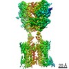
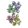
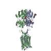
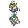
| Entry | Database: PDB / ID: 7lby | ||||||||||||
|---|---|---|---|---|---|---|---|---|---|---|---|---|---|
| Title | Bacterial cellulose synthase BcsB with polyalanine BcsA model | ||||||||||||
 Components Components |
| ||||||||||||
 Keywords Keywords | TRANSFERASE / bacterial cellulose / synthase / structural subunit / BIOSYNTHETIC PROTEIN | ||||||||||||
| Function / homology |  Function and homology information Function and homology informationbacterial cellulose biosynthetic process / Biofilm formation / cellulose synthase (UDP-forming) / cellulose synthase (UDP-forming) activity / cellulose biosynthetic process / UDP-alpha-D-glucose metabolic process / hexosyltransferase activity / cyclic-di-GMP binding / plasma membrane Similarity search - Function | ||||||||||||
| Biological species |  | ||||||||||||
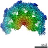
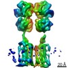
| Method | ELECTRON MICROSCOPY / single particle reconstruction / cryo EM / Resolution: 4.2 Å | ||||||||||||
 Authors Authors | Acheson, J.F. / Zimmer, J. | ||||||||||||
| Funding support |  United States, 3items United States, 3items
| ||||||||||||
 Citation Citation | Journal: Nat Struct Mol Biol / Year: 2021 Title: Molecular organization of the E. coli cellulose synthase macrocomplex. Authors: Justin F Acheson / Ruoya Ho / Nicolette F Goularte / Lynette Cegelski / Jochen Zimmer / Abstract: Cellulose is frequently found in communities of sessile bacteria called biofilms. Escherichia coli and other enterobacteriaceae modify cellulose with phosphoethanolamine (pEtN) to promote host tissue ...Cellulose is frequently found in communities of sessile bacteria called biofilms. Escherichia coli and other enterobacteriaceae modify cellulose with phosphoethanolamine (pEtN) to promote host tissue adhesion. The E. coli pEtN cellulose biosynthesis machinery contains the catalytic BcsA-B complex that synthesizes and secretes cellulose, in addition to five other subunits. The membrane-anchored periplasmic BcsG subunit catalyzes pEtN modification. Here we present the structure of the roughly 1 MDa E. coli Bcs complex, consisting of one BcsA enzyme associated with six copies of BcsB, determined by single-particle cryo-electron microscopy. BcsB homo-oligomerizes primarily through interactions between its carbohydrate-binding domains as well as intermolecular beta-sheet formation. The BcsB hexamer creates a half spiral whose open side accommodates two BcsG subunits, directly adjacent to BcsA's periplasmic channel exit. The cytosolic BcsE and BcsQ subunits associate with BcsA's regulatory PilZ domain. The macrocomplex is a fascinating example of cellulose synthase specification. #1: Journal: Nat.Struct.Mol.Biol. / Year: 2021Title: Molecular Organization of the E. coli Cellulose Synthase Macrocomplex Authors: Acheson, J.F. / Ho, R. / Goularte, N.F. / Cegelski, L. / Zimmer, J. | ||||||||||||
| History |
|
- Structure visualization
| Movie |
Movie viewer |
|---|---|
| Structure viewer | Molecule: MolmilJmol/JSmol |
- Downloads & links
-Download
| PDBx/mmCIF format | 7lby.cif.gz | 260 KB | Display | PDBx/mmCIF format |
|---|---|---|---|---|
| PDB format | pdb7lby.ent.gz | 189.6 KB | Display | PDB format |
| PDBx/mmJSON format | 7lby.json.gz | Tree view | PDBx/mmJSON format | |
| Others |  Other downloads Other downloads |
-Validation report
| Arichive directory | https://data.pdbj.org/pub/pdb/validation_reports/lb/7lbyftp://data.pdbj.org/pub/pdb/validation_reports/lb/7lby | HTTPS FTP |
|---|
-Related structure data
| Related structure data |  23267MC 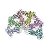 7l2zC M: map data used to model this data C: citing same article ( |
|---|---|
| Similar structure data | |
| EM raw data | EMPIAR-10627 (Title: Poly-alanine backbone model of E. coli BcsA bound to BcsB Data size: 3.2 TB Data #1: Unaligned multi-frame micrographs [micrographs - multiframe] Data #2: Unaligned movie frames [micrographs - multiframe] / Data #3: Unaligned movie frames [micrographs - multiframe] / Data #4: Unaligned movie frames [micrographs - multiframe] / Data #5: Unaligned movie frames [micrographs - multiframe]) |


-Links
 PDBj
PDBj

- Assembly
| Deposited unit | 
|
|---|---|
| 1 |
|
-Components
| #1: Protein | Mass: 101671.250 Da / Num. of mol.: 1 Source method: isolated from a genetically manipulated source Source: (gene. exp.) Strain: K12 / Gene: bcsA, yhjO, yhjP, b3533, JW5665 / Plasmid: pETDuetEcBcs_A-12His_nSS-Strep-B_Adra-6His / Production host: References: UniProt: P37653, cellulose synthase (UDP-forming) |
|---|---|
| #2: Protein | Mass: 86101.289 Da / Num. of mol.: 1 Source method: isolated from a genetically manipulated source Source: (gene. exp.) Strain: K12 / Gene: bcsB, yhjN, b3532, JW3500 / Plasmid: pETDuetEcBcs_A-12His_nSS-Strep-B_Adra-6His / Production host: |
| Has protein modification | Y |
-Experimental details
-Experiment
| Experiment | Method: ELECTRON MICROSCOPY |
|---|---|
| EM experiment | Aggregation state: PARTICLE / 3D reconstruction method: single particle reconstruction |
- Sample preparation
| Component | Name: Bacterial cellulose synthase BcsB with BcsA / Type: COMPLEX Details: BcsB monomer with BcsA refined using masked local refinement and signal subtraction from E coli BCS inner-membrane complex projections. Entity ID: all / Source: RECOMBINANT | ||||||||||||||||||||||||||||||||||||||||
|---|---|---|---|---|---|---|---|---|---|---|---|---|---|---|---|---|---|---|---|---|---|---|---|---|---|---|---|---|---|---|---|---|---|---|---|---|---|---|---|---|---|
| Molecular weight | Value: 0.180 MDa / Experimental value: NO | ||||||||||||||||||||||||||||||||||||||||
| Source (natural) | Organism: | ||||||||||||||||||||||||||||||||||||||||
| Source (recombinant) | Organism: | ||||||||||||||||||||||||||||||||||||||||
| Buffer solution | pH: 8 Details: All buffers made fresh from concentrated stock. A 10% LMNG 2% DMNG 2% CHS solution was made made in milliQ water without buffer. After rocking for one day at room temp the solution was ...Details: All buffers made fresh from concentrated stock. A 10% LMNG 2% DMNG 2% CHS solution was made made in milliQ water without buffer. After rocking for one day at room temp the solution was briefly sonicated to completely dissolve the CHS. | ||||||||||||||||||||||||||||||||||||||||
| Buffer component |
| ||||||||||||||||||||||||||||||||||||||||
| Specimen | Conc.: 0.4 mg/ml / Embedding applied: NO / Shadowing applied: NO / Staining applied: NO / Vitrification applied: YES | ||||||||||||||||||||||||||||||||||||||||
| Specimen support | Details: 2 drops amylamine added to the chamber / Grid material: COPPER / Grid mesh size: 300 divisions/in. / Grid type: C-flat-1.2/1.3 | ||||||||||||||||||||||||||||||||||||||||
| Vitrification | Instrument: FEI VITROBOT MARK IV / Cryogen name: ETHANE / Humidity: 100 % / Chamber temperature: 277 K Details: 3.5 uL applied to c-flat 1.2/1.3 300 mesh grids incubated for 30s then blotted using force 6 for 12 seconds |
- Electron microscopy imaging
| Experimental equipment |  Model: Titan Krios / Image courtesy: FEI Company |
|---|---|
| Microscopy | Model: FEI TITAN KRIOS |
| Electron gun | Electron source:  FIELD EMISSION GUN / Accelerating voltage: 300 kV / Illumination mode: FLOOD BEAM FIELD EMISSION GUN / Accelerating voltage: 300 kV / Illumination mode: FLOOD BEAM |
| Electron lens | Mode: BRIGHT FIELD / Nominal magnification: 81000 X / Nominal defocus max: -2500 nm / Nominal defocus min: -1000 nm / Cs: 2.7 mm |
| Specimen holder | Cryogen: NITROGEN / Specimen holder model: FEI TITAN KRIOS AUTOGRID HOLDER |
| Image recording | Electron dose: 51 e/Å2 / Film or detector model: GATAN K3 BIOQUANTUM (6k x 4k) / Num. of grids imaged: 1 / Num. of real images: 2964 / Details: movie mode 40 frames |
| EM imaging optics | Energyfilter name: GIF Bioquantum / Energyfilter slit width: 10 eV |
- Processing
| Software | Name: PHENIX / Version: 1.18_3845: / Classification: refinement | ||||||||||||||||||||||||||||||
|---|---|---|---|---|---|---|---|---|---|---|---|---|---|---|---|---|---|---|---|---|---|---|---|---|---|---|---|---|---|---|---|
| EM software |
| ||||||||||||||||||||||||||||||
| CTF correction | Type: PHASE FLIPPING AND AMPLITUDE CORRECTION | ||||||||||||||||||||||||||||||
| Particle selection | Num. of particles selected: 409611 | ||||||||||||||||||||||||||||||
| 3D reconstruction | Resolution: 4.2 Å / Resolution method: FSC 0.143 CUT-OFF / Num. of particles: 193152 / Symmetry type: POINT | ||||||||||||||||||||||||||||||
| Atomic model building | B value: 101.45 / Protocol: AB INITIO MODEL / Space: REAL / Target criteria: CC | ||||||||||||||||||||||||||||||
| Refine LS restraints |
|
