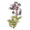
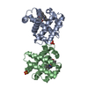
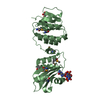
A: Bacterial cellulose synthesis subunit E hetero molecules
defined by author
Evidence: light scattering, SEC-MALS measurements in absence and presence of c-di-GMP have demonstrated that this construct remains monomeric in solution.
Type: DECTRIS EIGER X 16M / Detector: PIXEL / Date: Jun 3, 2017
Radiation
Protocol: SINGLE WAVELENGTH / Monochromatic (M) / Laue (L): M / Scattering type: x-ray
Radiation wavelength
ID
Wavelength (Å)
Relative weight
1
0.97911
1
2
0.97927
1
Reflection
Resolution: 2.2→50 Å / Num. obs: 66468 / % possible obs: 99.4 % / Redundancy: 9.13 % / Biso Wilson estimate: 50.37 Å2 / CC1/2: 0.998 / Rpim(I) all: 0.052 / Rrim(I) all: 0.217 / Net I/σ(I): 9.11
Reflection shell
Resolution: 2.2→2.33 Å / Mean I/σ(I) obs: 0.81 / Num. unique obs: 10635 / CC1/2: 0.458 / % possible all: 98.2
-
Processing
Software
Name
Version
Classification
BUSTER
2.10.3
refinement
PHENIX
AUTOBUILD
modelbuilding
BUCCANEER
modelbuilding
PHASER
v2.5.7
phasing
HKL2Map
Version2016
phasing
XDS
VERSIONNov1, 2016
dataprocessing
Refinement
Method to determine structure: SAD / Resolution: 2.2→49.73 Å / Cor.coef. Fo:Fc: 0.946 / Cor.coef. Fo:Fc free: 0.933 / SU R Cruickshank DPI: 0.221 / Cross valid method: THROUGHOUT / σ(F): 0 / SU R Blow DPI: 0.223 / SU Rfree Blow DPI: 0.182 / SU Rfree Cruickshank DPI: 0.183
Rfactor
Num. reflection
% reflection
Selection details
Rfree
0.238
1759
5.01 %
RANDOM
Rwork
0.205
-
-
-
obs
0.207
35118
99.6 %
-
Displacement parameters
Biso mean: 59.86 Å2
Baniso -1
Baniso -2
Baniso -3
1-
2.616 Å2
0 Å2
0 Å2
2-
-
2.616 Å2
0 Å2
3-
-
-
-5.2321 Å2
Refine analyze
Luzzati coordinate error obs: 0.34 Å
Refinement step
Cycle: 1 / Resolution: 2.2→49.73 Å
Protein
Nucleic acid
Ligand
Solvent
Total
Num. atoms
4091
0
58
150
4299
Refine LS restraints
Refine-ID
Type
Dev ideal
Number
Restraint function
Weight
X-RAY DIFFRACTION
t_bond_d
0.009
4272
HARMONIC
2
X-RAY DIFFRACTION
t_angle_deg
1.05
5840
HARMONIC
2
X-RAY DIFFRACTION
t_dihedral_angle_d
1517
SINUSOIDAL
2
X-RAY DIFFRACTION
t_incorr_chiral_ct
X-RAY DIFFRACTION
t_pseud_angle
X-RAY DIFFRACTION
t_trig_c_planes
112
HARMONIC
2
X-RAY DIFFRACTION
t_gen_planes
639
HARMONIC
5
X-RAY DIFFRACTION
t_it
4272
HARMONIC
20
X-RAY DIFFRACTION
t_nbd
X-RAY DIFFRACTION
t_omega_torsion
2.86
X-RAY DIFFRACTION
t_other_torsion
16.34
X-RAY DIFFRACTION
t_improper_torsion
X-RAY DIFFRACTION
t_chiral_improper_torsion
560
SEMIHARMONIC
5
X-RAY DIFFRACTION
t_sum_occupancies
X-RAY DIFFRACTION
t_utility_distance
X-RAY DIFFRACTION
t_utility_angle
X-RAY DIFFRACTION
t_utility_torsion
X-RAY DIFFRACTION
t_ideal_dist_contact
4866
SEMIHARMONIC
4
LS refinement shell
Resolution: 2.2→2.26 Å / Total num. of bins used: 18
Rfactor
Num. reflection
% reflection
Rfree
0.2496
141
5.02 %
Rwork
0.2528
2669
-
all
0.2526
2810
-
obs
-
-
99.33 %
Refinement TLS params.
Method: refined / Refine-ID: X-RAY DIFFRACTION
ID
L11 (°2)
L12 (°2)
L13 (°2)
L22 (°2)
L23 (°2)
L33 (°2)
S11 (Å °)
S12 (Å °)
S13 (Å °)
S21 (Å °)
S22 (Å °)
S23 (Å °)
S31 (Å °)
S32 (Å °)
S33 (Å °)
T11 (Å2)
T12 (Å2)
T13 (Å2)
T22 (Å2)
T23 (Å2)
T33 (Å2)
Origin x (Å)
Origin y (Å)
Origin z (Å)
1
0.3051
0.7311
-0.2464
2.8716
-0.6359
0.4127
-0.0311
-0.0215
-0.0483
0.1317
0.0345
-0.0401
0.0349
0.0302
-0.0034
-0.2274
0.0296
-0.0231
-0.2646
0.0081
0.3588
44.7542
134.278
126.83
2
0.3312
-0.789
0.2332
3.9674
-0.6954
0.2316
0.0155
0.1122
0.025
-0.0034
-0.0394
-0.2078
0.0218
0.0616
0.0238
-0.2969
-0.0089
-0.0014
-0.2985
-0.003
0.3876
42.4709
87.7611
111.135
Refinement TLS group
ID
Refine-ID
Refine TLS-ID
Selection details
1
X-RAY DIFFRACTION
1
{ A|* }
2
X-RAY DIFFRACTION
2
{ B|* }
+
About Yorodumi
-
News
-
Feb 9, 2022. New format data for meta-information of EMDB entries
New format data for meta-information of EMDB entries
Version 3 of the EMDB header file is now the official format.
The previous official version 1.9 will be removed from the archive.
In the structure databanks used in Yorodumi, some data are registered as the other names, "COVID-19 virus" and "2019-nCoV". Here are the details of the virus and the list of structure data.
Jan 31, 2019. EMDB accession codes are about to change! (news from PDBe EMDB page)
EMDB accession codes are about to change! (news from PDBe EMDB page)
The allocation of 4 digits for EMDB accession codes will soon come to an end. Whilst these codes will remain in use, new EMDB accession codes will include an additional digit and will expand incrementally as the available range of codes is exhausted. The current 4-digit format prefixed with “EMD-” (i.e. EMD-XXXX) will advance to a 5-digit format (i.e. EMD-XXXXX), and so on. It is currently estimated that the 4-digit codes will be depleted around Spring 2019, at which point the 5-digit format will come into force.
The EM Navigator/Yorodumi systems omit the EMD- prefix.
Related info.:Q: What is EMD? / ID/Accession-code notation in Yorodumi/EM Navigator
Yorodumi is a browser for structure data from EMDB, PDB, SASBDB, etc.
This page is also the successor to EM Navigator detail page, and also detail information page/front-end page for Omokage search.
The word "yorodu" (or yorozu) is an old Japanese word meaning "ten thousand". "mi" (miru) is to see.
Related info.:EMDB / PDB / SASBDB / Comparison of 3 databanks / Yorodumi Search / Aug 31, 2016. New EM Navigator & Yorodumi / Yorodumi Papers / Jmol/JSmol / Function and homology information / Changes in new EM Navigator and Yorodumi
 Movie
Movie Controller

 Yorodumi
 Open data
 Basic information
 Components
 Keywords
 Function and homology information
 X-RAY DIFFRACTION /
  Authors
 Citation
 Structure visualization
 Downloads & links
 Other downloads
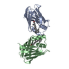


 PDBj
PDBj
 Assembly


 Mass: 92.094 Da / Num. of mol.: 2 / Source method: obtained synthetically / Formula: C3H8O3 / Feature type: SUBJECT OF INVESTIGATION
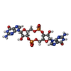
 Mass: 690.411 Da / Num. of mol.: 1 / Source method: obtained synthetically / Formula: C20H24N10O14P2 / Feature type: SUBJECT OF INVESTIGATION
 Mass: 18.015 Da / Num. of mol.: 150 / Source method: isolated from a natural source / Formula: H2O
 Sample preparation
 / Beamline: PROXIMA 1 / Wavelength: 0.97911, 0.97927
 Processing