 Movie
Movie Controller

[English] 日本語
 Yorodumi
- PDB-4j29: Crystal Structure of Engineered Protein. Northeast Structural Gen... -
+ Open data

- Basic information
| Entry | Database: PDB / ID: 4j29 | ||||||
|---|---|---|---|---|---|---|---|
| Title | Crystal Structure of Engineered Protein. Northeast Structural Genomics Consortium Target OR258. | ||||||
 Components Components | Engineered Protein OR258 | ||||||
 Keywords Keywords | Structural Genomics / Unknown Function / PSI-Biology / Protein Structure Initiative / NESG / OR258 / Northeast Structural Genomics Consortium | ||||||
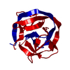
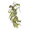
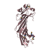
| Function / homology | Rossmann fold - #11230 / Rossmann fold / 3-Layer(aba) Sandwich / Alpha Beta Function and homology information Function and homology information | ||||||
| Biological species | synthetic construct (others) | ||||||
| Method |  X-RAY DIFFRACTION / SYNCHROTRON / SAD / Resolution: 2.1 Å X-RAY DIFFRACTION / SYNCHROTRON / SAD / Resolution: 2.1 Å | ||||||
 Authors Authors | Vorobiev, S. / Su, M. / Koga, R. / Seetharaman, J. / Koga, N. / Mao, L. / Xiao, R. / Kohan, E. / Castelllanos, J. / Everett, J.K. ...Vorobiev, S. / Su, M. / Koga, R. / Seetharaman, J. / Koga, N. / Mao, L. / Xiao, R. / Kohan, E. / Castelllanos, J. / Everett, J.K. / Acton, T.B. / Baker, D. / Montelione, G.T. / Tong, L. / Hunt, J.F. / Northeast Structural Genomics Consortium (NESG) | ||||||
 Citation Citation | Journal: To be Published Title: Crystal Structure of Engineered Protein OR258. Authors: Vorobiev, S. / Su, M. / Koga, R. / Seetharaman, J. / Koga, N. / Mao, L. / Xiao, R. / Kohan, E. / Castelllanos, J. / Everett, J.K. / Acton, T.B. / Baker, D. / Montelione, G.T. / Tong, L. / Hunt, J.F. | ||||||
| History |
|
- Structure visualization
| Structure viewer | Molecule: MolmilJmol/JSmol |
|---|
- Downloads & links
-Download
| PDBx/mmCIF format | 4j29.cif.gz | 64.8 KB | Display | PDBx/mmCIF format |
|---|---|---|---|---|
| PDB format | pdb4j29.ent.gz | 48 KB | Display | PDB format |
| PDBx/mmJSON format | 4j29.json.gz | Tree view | PDBx/mmJSON format | |
| Others |  Other downloads Other downloads |
-Validation report
| Arichive directory | https://data.pdbj.org/pub/pdb/validation_reports/j2/4j29ftp://data.pdbj.org/pub/pdb/validation_reports/j2/4j29 | HTTPS FTP |
|---|
-Related structure data
| Similar structure data | |
|---|---|
| Other databases |


-Links
 PDBj
PDBj- Assembly
| Deposited unit | 
| ||||||||
|---|---|---|---|---|---|---|---|---|---|
| 1 | 
| ||||||||
| Unit cell |
| ||||||||
| Components on special symmetry positions |
|
-Components
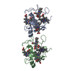
| #1: Protein | Mass: 15862.859 Da / Num. of mol.: 1 Source method: isolated from a genetically manipulated source Source: (gene. exp.) synthetic construct (others) / Plasmid: pET29b+, OR258-29.1 / Production host:  |
|---|---|
| #2: Water | ChemComp-HOH /  Mass: 18.015 Da / Num. of mol.: 16 / Source method: isolated from a natural source / Formula: H2O Mass: 18.015 Da / Num. of mol.: 16 / Source method: isolated from a natural source / Formula: H2O |
| Has protein modification | Y |
-Experimental details
-Experiment
| Experiment | Method: X-RAY DIFFRACTION / Number of used crystals: 1 |
|---|
- Sample preparation
| Crystal | Density Matthews: 1.76 Å3/Da / Density % sol: 30.07 % |
|---|---|
| Crystal grow | Method: vapor diffusion, hanging drop / pH: 7.5 Details: 100mM NaCl, 5mM DTT, 0.02% NaN3, 10mM Tris-HCl pH 7.5, VAPOR DIFFUSION, HANGING DROP |
-Data collection
| Diffraction | Mean temperature: 100 K |
|---|---|
| Diffraction source | Source: SYNCHROTRON / Site: APS  / Beamline: 24-ID-E / Wavelength: 0.97921 Å / Beamline: 24-ID-E / Wavelength: 0.97921 Å |
| Detector | Type: ADSC QUANTUM 315 / Detector: CCD / Date: Dec 6, 2012 |
| Radiation | Protocol: SINGLE WAVELENGTH / Monochromatic (M) / Laue (L): M / Scattering type: x-ray |
| Radiation wavelength | Wavelength: 0.97921 Å / Relative weight: 1 |
| Reflection | Resolution: 2.1→50 Å / Num. all: 12677 / Num. obs: 12646 / % possible obs: 99.8 % / Observed criterion σ(F): 0 / Observed criterion σ(I): 0 / Redundancy: 27.6 % / Biso Wilson estimate: 36.16 Å2 / Rmerge(I) obs: 0.151 / Net I/σ(I): 22.3 |
| Reflection shell | Resolution: 2.1→2.21 Å / Redundancy: 28.5 % / Rmerge(I) obs: 1.87 / Mean I/σ(I) obs: 2.3 / Num. unique all: 1663 / % possible all: 99.9 |
- Processing
| Software |
| ||||||||||||||||||||||||||||||||||||||||
|---|---|---|---|---|---|---|---|---|---|---|---|---|---|---|---|---|---|---|---|---|---|---|---|---|---|---|---|---|---|---|---|---|---|---|---|---|---|---|---|---|---|
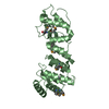
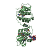
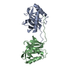
| Refinement | Method to determine structure: SAD / Resolution: 2.1→23.885 Å / Occupancy max: 1 / Occupancy min: 0.88 / SU ML: 0.69 / Cross valid method: THROUGHOUT / σ(F): 1.35 / Phase error: 25.44 / Stereochemistry target values: ML Details: IN THE CRYSTAL STRUCTURE OR258 FORMS DIMER WITH CRYSTAL SYMMETRY MATE (-X+1,-Y+1,Z) BY DOMAIN SWAPPING.
| ||||||||||||||||||||||||||||||||||||||||
| Solvent computation | Shrinkage radii: 0.86 Å / VDW probe radii: 1.1 Å / Solvent model: FLAT BULK SOLVENT MODEL / Bsol: 62.951 Å2 / ksol: 0.38 e/Å3 | ||||||||||||||||||||||||||||||||||||||||
| Displacement parameters | Biso max: 104.57 Å2 / Biso mean: 46.611 Å2 / Biso min: 20.24 Å2
| ||||||||||||||||||||||||||||||||||||||||
| Refinement step | Cycle: LAST / Resolution: 2.1→23.885 Å
| ||||||||||||||||||||||||||||||||||||||||
| Refine LS restraints |
| ||||||||||||||||||||||||||||||||||||||||
| LS refinement shell | Refine-ID: X-RAY DIFFRACTION / Total num. of bins used: 4 / % reflection obs: 100 %
| ||||||||||||||||||||||||||||||||||||||||
| Refinement TLS params. | Method: refined / Origin x: 34.5346 Å / Origin y: 25.6132 Å / Origin z: 12.4845 Å
| ||||||||||||||||||||||||||||||||||||||||
| Refinement TLS group | Selection details: chain A |
