 Movie
Movie Controller

[English] 日本語
 Yorodumi
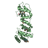
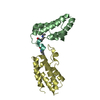
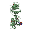
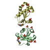
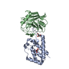
- PDB-1mi8: 2.0 Angstrom crystal structure of a DnaB intein from Synechocysti... -
+ Open data

- Basic information
| Entry | Database: PDB / ID: 1mi8 | ||||||
|---|---|---|---|---|---|---|---|
| Title | 2.0 Angstrom crystal structure of a DnaB intein from Synechocystis sp. PCC 6803 | ||||||
 Components Components | DnaB intein | ||||||
 Keywords Keywords | HYDROLASE / all beta-strands | ||||||
| Function / homology |  Function and homology information Function and homology informationprimosome complex / intein-mediated protein splicing / DNA replication, synthesis of primer / DNA 5'-3' helicase / DNA helicase activity / endonuclease activity / 5'-3' DNA helicase activity / Hydrolases; Acting on ester bonds / DNA replication / ATP hydrolysis activity ...primosome complex / intein-mediated protein splicing / DNA replication, synthesis of primer / DNA 5'-3' helicase / DNA helicase activity / endonuclease activity / 5'-3' DNA helicase activity / Hydrolases; Acting on ester bonds / DNA replication / ATP hydrolysis activity / DNA binding / ATP binding / cytosol Similarity search - Function | ||||||
| Biological species |  | ||||||
| Method |  X-RAY DIFFRACTION / MOLECULAR REPLACEMENT / Resolution: 2 Å X-RAY DIFFRACTION / MOLECULAR REPLACEMENT / Resolution: 2 Å | ||||||
 Authors Authors | Ding, Y. / Chen, X. / Ferrandon, S. / Xu, M. / Rao, Z. | ||||||
 Citation Citation | Journal: J.Biol.Chem. / Year: 2003 Title: Crystal structure of mini-intein reveals a conserved catalytic module involved in side chain cyclization of asparagine during protein splicing Authors: Ding, Y. / Xu, M.Q. / Ghosh, I. / Chen, X. / Ferrandon, S. / Lesage, G. / Rao, Z. | ||||||
| History |
|
- Structure visualization
| Structure viewer | Molecule: MolmilJmol/JSmol |
|---|
- Downloads & links
-Download
| PDBx/mmCIF format | 1mi8.cif.gz | 40.2 KB | Display | PDBx/mmCIF format |
|---|---|---|---|---|
| PDB format | pdb1mi8.ent.gz | 27.9 KB | Display | PDB format |
| PDBx/mmJSON format | 1mi8.json.gz | Tree view | PDBx/mmJSON format | |
| Others |  Other downloads Other downloads |
-Validation report
| Arichive directory | https://data.pdbj.org/pub/pdb/validation_reports/mi/1mi8ftp://data.pdbj.org/pub/pdb/validation_reports/mi/1mi8 | HTTPS FTP |
|---|
-Related structure data
| Similar structure data |
|---|


-Links
 PDBj
PDBj


- Assembly
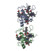
| Deposited unit | 
| ||||||||
|---|---|---|---|---|---|---|---|---|---|
| 1 | 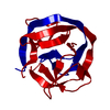
| ||||||||
| Unit cell |
| ||||||||
| Details | The biological assembly is a dimer generated from the monomer in the asymmetric unit |
-Components
| #1: Protein | Mass: 17600.168 Da / Num. of mol.: 1 Source method: isolated from a genetically manipulated source Details: TETHERED DIMER LINKED BY LESSSLQLSPEIEKLSQ / Source: (gene. exp.) References: UniProt: Q55418, Hydrolases; Acting on acid anhydrides; In phosphorus-containing anhydrides |
|---|---|
| #2: Water | ChemComp-HOH /  Mass: 18.015 Da / Num. of mol.: 50 / Source method: isolated from a natural source / Formula: H2O Mass: 18.015 Da / Num. of mol.: 50 / Source method: isolated from a natural source / Formula: H2O |
-Experimental details
-Experiment
| Experiment | Method: X-RAY DIFFRACTION / Number of used crystals: 1 |
|---|
- Sample preparation
| Crystal | Density Matthews: 1.95 Å3/Da / Density % sol: 36.92 % | ||||||||||||||||||||||||
|---|---|---|---|---|---|---|---|---|---|---|---|---|---|---|---|---|---|---|---|---|---|---|---|---|---|
| Crystal grow | Temperature: 293 K / Method: vapor diffusion, hanging drop / pH: 7.8 Details: PEG4000, Tris-HCl, pH 7.8, VAPOR DIFFUSION, HANGING DROP, temperature 293K | ||||||||||||||||||||||||
| Crystal grow | *PLUS Method: vapor diffusion, hanging drop / PH range low: 8 / PH range high: 7.5 | ||||||||||||||||||||||||
| Components of the solutions | *PLUS
|
-Data collection
| Diffraction | Mean temperature: 100 K |
|---|---|
| Diffraction source | Source: ROTATING ANODE / Type: RIGAKU RU200 / Wavelength: 1.5418 Å |
| Detector | Type: MARRESEARCH / Detector: IMAGE PLATE / Date: May 8, 2002 |
| Radiation | Monochromator: osmic mirror / Protocol: SINGLE WAVELENGTH / Monochromatic (M) / Laue (L): M / Scattering type: x-ray |
| Radiation wavelength | Wavelength: 1.5418 Å / Relative weight: 1 |
| Reflection | Resolution: 2→50 Å / Num. obs: 9307 / % possible obs: 99.9 % / Observed criterion σ(F): 1 / Observed criterion σ(I): 1 |
| Reflection shell | Resolution: 2→2.07 Å / % possible all: 75.5 |
| Reflection | *PLUS Lowest resolution: 40 Å / % possible obs: 97.6 % / Num. measured all: 107903 / Rmerge(I) obs: 0.046 |
| Reflection shell | *PLUS Highest resolution: 2 Å / % possible obs: 75.5 % / Rmerge(I) obs: 0.262 / Mean I/σ(I) obs: 7.5 |
- Processing
| Software |
| ||||||||||||||||||||
|---|---|---|---|---|---|---|---|---|---|---|---|---|---|---|---|---|---|---|---|---|---|
| Refinement | Method to determine structure: MOLECULAR REPLACEMENT / Resolution: 2→20 Å / σ(F): 2 / Stereochemistry target values: Engh & Huber
| ||||||||||||||||||||
| Refinement step | Cycle: LAST / Resolution: 2→20 Å
| ||||||||||||||||||||
| Refine LS restraints |
| ||||||||||||||||||||
| Refinement | *PLUS Lowest resolution: 40 Å / % reflection Rfree: 10 % / Rfactor Rwork: 0.21 | ||||||||||||||||||||
| Solvent computation | *PLUS | ||||||||||||||||||||
| Displacement parameters | *PLUS |
