 Movie
Movie Controller


+ Open data

- Basic information
| Entry | Database: PDB / ID: 7jw3 | ||||||
|---|---|---|---|---|---|---|---|
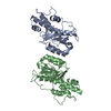
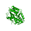
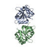
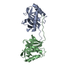
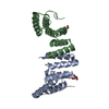
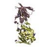
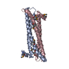
| Title | Crystal structure of Aedes aegypti Nibbler NTD domain | ||||||
 Components Components | Exonuclease mut-7 homolog | ||||||
 Keywords Keywords | RNA BINDING PROTEIN / Nibbler / exoribonuclease / microRNA trimming / piRNA trimming / HEAT | ||||||
| Function / homology |  Function and homology information Function and homology informationnucleobase-containing compound metabolic process / 3'-5' exonuclease activity / nucleic acid binding / Hydrolases; Acting on ester bonds / metal ion binding Similarity search - Function | ||||||
| Biological species |  | ||||||
| Method |  X-RAY DIFFRACTION / SYNCHROTRON / SAD / Resolution: 3.05 Å X-RAY DIFFRACTION / SYNCHROTRON / SAD / Resolution: 3.05 Å | ||||||
 Authors Authors | Xie, W. / Sowemimo, I. / Hayashi, R. / Wang, J. / Brennecke, J. / Ameres, S.L. / Patel, D.J. | ||||||
 Citation Citation | Journal: Proc.Natl.Acad.Sci.USA / Year: 2020 Title: Structure-function analysis of microRNA 3'-end trimming by Nibbler. Authors: Xie, W. / Sowemimo, I. / Hayashi, R. / Wang, J. / Burkard, T.R. / Brennecke, J. / Ameres, S.L. / Patel, D.J. | ||||||
| History |
|
- Structure visualization
| Structure viewer | Molecule: MolmilJmol/JSmol |
|---|
- Downloads & links
-Download
| PDBx/mmCIF format | 7jw3.cif.gz | 170 KB | Display | PDBx/mmCIF format |
|---|---|---|---|---|
| PDB format | pdb7jw3.ent.gz | 131.6 KB | Display | PDB format |
| PDBx/mmJSON format | 7jw3.json.gz | Tree view | PDBx/mmJSON format | |
| Others |  Other downloads Other downloads |
-Validation report
| Arichive directory | https://data.pdbj.org/pub/pdb/validation_reports/jw/7jw3ftp://data.pdbj.org/pub/pdb/validation_reports/jw/7jw3 | HTTPS FTP |
|---|
-Related structure data


-Links
 PDBj
PDBj
- Assembly
| Deposited unit | 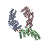
| ||||||||||||
|---|---|---|---|---|---|---|---|---|---|---|---|---|---|
| 1 | 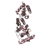
| ||||||||||||
| 2 | 
| ||||||||||||
| 3 | 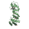
| ||||||||||||
| Unit cell |
|
-Components
| #1: Protein | Mass: 45623.625 Da / Num. of mol.: 3 Source method: isolated from a genetically manipulated source Source: (gene. exp.)  References: UniProt: Q179T2, Hydrolases; Acting on ester bonds Has ligand of interest | N | Has protein modification | Y | |
|---|
-Experimental details
-Experiment
| Experiment | Method: X-RAY DIFFRACTION / Number of used crystals: 1 |
|---|
- Sample preparation
| Crystal grow | Temperature: 293 K / Method: vapor diffusion, hanging drop / pH: 7.5 Details: 0.2 M sodium tartrate, 0.1 M HEPES, pH 7.0, 20% PEG3350 |
|---|
-Data collection
| Diffraction | Mean temperature: 100 K / Serial crystal experiment: N |
|---|---|
| Diffraction source | Source: SYNCHROTRON / Site: APS  / Beamline: 24-ID-C / Wavelength: 0.9792 Å / Beamline: 24-ID-C / Wavelength: 0.9792 Å |
| Detector | Type: DECTRIS PILATUS3 S 6M / Detector: PIXEL / Date: Mar 8, 2017 |
| Radiation | Protocol: SINGLE WAVELENGTH / Monochromatic (M) / Laue (L): M / Scattering type: x-ray |
| Radiation wavelength | Wavelength: 0.9792 Å / Relative weight: 1 |
| Reflection | Resolution: 3.05→30 Å / Num. obs: 21285 / % possible obs: 99.73 % / Redundancy: 20.2 % / Biso Wilson estimate: 114.23 Å2 / CC1/2: 0.999 / CC star: 1 / Rmerge(I) obs: 0.1599 / Rpim(I) all: 0.03625 / Rrim(I) all: 0.164 / Net I/σ(I): 18.12 |
| Reflection shell | Resolution: 3.05→3.159 Å / Redundancy: 20.1 % / Rmerge(I) obs: 3.551 / Mean I/σ(I) obs: 1.05 / Num. unique obs: 2058 / CC1/2: 0.524 / CC star: 0.829 / Rpim(I) all: 0.8052 / Rrim(I) all: 3.642 / % possible all: 99.28 |
- Processing
| Software |
| ||||||||||||||||||||||||||||||||||||||||||||||||||||||||
|---|---|---|---|---|---|---|---|---|---|---|---|---|---|---|---|---|---|---|---|---|---|---|---|---|---|---|---|---|---|---|---|---|---|---|---|---|---|---|---|---|---|---|---|---|---|---|---|---|---|---|---|---|---|---|---|---|---|
| Refinement | Method to determine structure: SAD / Resolution: 3.05→29.21 Å / SU ML: 0.4802 / Cross valid method: FREE R-VALUE / σ(F): 1.35 / Phase error: 33.5035 Stereochemistry target values: GeoStd + Monomer Library + CDL v1.2
| ||||||||||||||||||||||||||||||||||||||||||||||||||||||||
| Solvent computation | Shrinkage radii: 0.9 Å / VDW probe radii: 1.11 Å / Solvent model: FLAT BULK SOLVENT MODEL | ||||||||||||||||||||||||||||||||||||||||||||||||||||||||
| Displacement parameters | Biso mean: 125.41 Å2 | ||||||||||||||||||||||||||||||||||||||||||||||||||||||||
| Refinement step | Cycle: LAST / Resolution: 3.05→29.21 Å
| ||||||||||||||||||||||||||||||||||||||||||||||||||||||||
| Refine LS restraints |
| ||||||||||||||||||||||||||||||||||||||||||||||||||||||||
| LS refinement shell |
|
