 Movie
Movie Controller

[English] 日本語
 Yorodumi
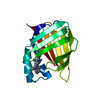
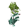
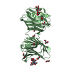
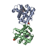
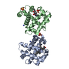
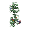
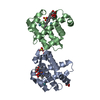
- PDB-1cbr: CRYSTAL STRUCTURE OF CELLULAR RETINOIC-ACID-BINDING PROTEINS I AN... -
+ Open data

- Basic information
| Entry | Database: PDB / ID: 1cbr | ||||||
|---|---|---|---|---|---|---|---|
| Title | CRYSTAL STRUCTURE OF CELLULAR RETINOIC-ACID-BINDING PROTEINS I AND II IN COMPLEX WITH ALL-TRANS-RETINOIC ACID AND A SYNTHETIC RETINOID | ||||||
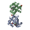
 Components Components | CELLULAR RETINOIC ACID BINDING PROTEIN TYPE I | ||||||
 Keywords Keywords | RETINOIC-ACID TRANSPORT | ||||||
| Function / homology |  Function and homology information Function and homology informationRA biosynthesis pathway / retinoid binding / retinoic acid binding / retinal binding / retinol binding / fatty acid transport / fatty acid binding / nucleus / cytoplasm / cytosol Similarity search - Function | ||||||
| Biological species |  | ||||||
| Method |  X-RAY DIFFRACTION / Resolution: 2.9 Å X-RAY DIFFRACTION / Resolution: 2.9 Å | ||||||
 Authors Authors | Kleywegt, G.J. / Bergfors, T. / Jones, T.A. | ||||||
 Citation Citation | Journal: Structure / Year: 1994 Title: Crystal structures of cellular retinoic acid binding proteins I and II in complex with all-trans-retinoic acid and a synthetic retinoid. Authors: Kleywegt, G.J. / Bergfors, T. / Senn, H. / Le Motte, P. / Gsell, B. / Shudo, K. / Jones, T.A. #1: Journal: Adv.Protein Chem. / Year: 1994Title: Lipid-Binding Proteins: A Family of Fatty Acid and Retinoid Transport Proteins Authors: Banaszak, L. / Winter, N. / Xu, Z. / Bernlohr, D.A. / Cowan, S.W. #2: Journal: Acta Crystallogr.,Sect.D / Year: 1994Title: Crystallisation and Preliminary X-Ray Analysis of Recombinant Bovine Cellular Retinoic Acid-Binding Protein Authors: Bergfors, T. / Kleywegt, G.J. / Jones, T.A. #3: Journal: J.Mol.Biol. / Year: 1993Title: Crystallographic Studies on a Family of Lipophilic Transport Proteins. Refinement of P2 Myelin Protein and the Structure Determination and Refinement of Cellular Retinol-Binding Protein in ...Title: Crystallographic Studies on a Family of Lipophilic Transport Proteins. Refinement of P2 Myelin Protein and the Structure Determination and Refinement of Cellular Retinol-Binding Protein in Complex with All-Trans-Retinol Authors: Cowan, S.W. / Newcomer, M.E. / Jones, T.A. #4: Journal: Embo J. / Year: 1988Title: The Three-Dimensional Structure of P2 Myelin Protein Authors: Jones, T.A. / Bergfors, T. / Sedzik, J. / Unge, T. | ||||||
| History |
|
- Structure visualization
| Structure viewer | Molecule: MolmilJmol/JSmol |
|---|
- Downloads & links
-Download
| PDBx/mmCIF format | 1cbr.cif.gz | 62.4 KB | Display | PDBx/mmCIF format |
|---|---|---|---|---|
| PDB format | pdb1cbr.ent.gz | 47.2 KB | Display | PDB format |
| PDBx/mmJSON format | 1cbr.json.gz | Tree view | PDBx/mmJSON format | |
| Others |  Other downloads Other downloads |
-Validation report
| Arichive directory | https://data.pdbj.org/pub/pdb/validation_reports/cb/1cbrftp://data.pdbj.org/pub/pdb/validation_reports/cb/1cbr | HTTPS FTP |
|---|
-Related structure data


-Links
 PDBj
PDBj

- Assembly
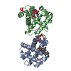
| Deposited unit | 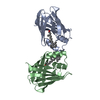
| ||||||||
|---|---|---|---|---|---|---|---|---|---|
| 1 |
| ||||||||
| Unit cell |
| ||||||||
| Noncrystallographic symmetry (NCS) | NCS oper: (Code: given Matrix: (0.781496, -0.392775, -0.484758), Vector: Details | MTRIX THE TRANSFORMATIONS PRESENTED ON MTRIX RECORDS BELOW DESCRIBE NON-CRYSTALLOGRAPHIC RELATIONSHIPS AMONG THE VARIOUS DOMAINS IN THIS ENTRY. APPLYING THE APPROPRIATE MTRIX TRANSFORMATION TO THE RESIDUES LISTED FIRST WILL YIELD APPROXIMATE COORDINATES FOR THE RESIDUES LISTED SECOND. APPLIED TO TRANSFORMED TO MTRIX RESIDUES RESIDUES RMSD M1 A 1 .. A 136 B 1 .. B 136 0.0 BECAUSE OF THE STRICT NON-CRYSTALLOGRAPHIC SYMMETRY USED IN REFINEMENT, THE TRANSFORMATION DESCRIBED, IN FACT, YIELDS EXACT COORDINATES FOR CHAIN B WHEN APPLIED TO CHAIN A. | |
-Components
| #1: Protein | Mass: 15480.392 Da / Num. of mol.: 2 Source method: isolated from a genetically manipulated source Source: (gene. exp.)  #2: Chemical | 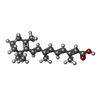  Mass: 300.435 Da / Num. of mol.: 2 / Source method: obtained synthetically / Formula: C20H28O2 Mass: 300.435 Da / Num. of mol.: 2 / Source method: obtained synthetically / Formula: C20H28O2#3: Water | ChemComp-HOH / |  Mass: 18.015 Da / Num. of mol.: 28 / Source method: isolated from a natural source / Formula: H2O Mass: 18.015 Da / Num. of mol.: 28 / Source method: isolated from a natural source / Formula: H2O |
|---|
-Experimental details
-Experiment
| Experiment | Method: X-RAY DIFFRACTION |
|---|
- Sample preparation
| Crystal | Density Matthews: 2.81 Å3/Da / Density % sol: 56.25 % | ||||||||||||||||||||||||||||||||||||||||||||||||||||||
|---|---|---|---|---|---|---|---|---|---|---|---|---|---|---|---|---|---|---|---|---|---|---|---|---|---|---|---|---|---|---|---|---|---|---|---|---|---|---|---|---|---|---|---|---|---|---|---|---|---|---|---|---|---|---|---|
| Crystal grow | *PLUS Temperature: 277 K / pH: 8 / Method: vapor diffusion, sitting dropDetails: Bergfors, T., (1994) Acta Crystallogr.,Sect.D, 50, 370. | ||||||||||||||||||||||||||||||||||||||||||||||||||||||
| Components of the solutions | *PLUS
|
-Data collection
| Radiation | Scattering type: x-ray |
|---|---|
| Radiation wavelength | Relative weight: 1 |
| Reflection | Num. obs: 7118 / Observed criterion σ(I): 3 |
| Reflection | *PLUS Highest resolution: 2.9 Å / Lowest resolution: 30 Å / % possible obs: 94.2 % / Num. measured all: 16898 / Rmerge(I) obs: 0.086 |
| Reflection shell | *PLUS Highest resolution: 2.9 Å / Lowest resolution: 2.98 Å |
- Processing
| Software |
| ||||||||||||||||||||||||||||||||||||||||||||||||||||||||||||
|---|---|---|---|---|---|---|---|---|---|---|---|---|---|---|---|---|---|---|---|---|---|---|---|---|---|---|---|---|---|---|---|---|---|---|---|---|---|---|---|---|---|---|---|---|---|---|---|---|---|---|---|---|---|---|---|---|---|---|---|---|---|
| Refinement | Resolution: 2.9→8 Å / σ(F): 2 Details: THE LIGAND FOR THIS ENTRY WAS TAKEN FROM ENTRY 1CBS AND ADJUSTED TO THE DENSITY AS A RIGID BODY BY X-PLOR. THIS HAS INTRODUCED A SHORT CONTACT (2.2 A) BETWEEN AN OXYGEN OF THE LIGAND AND THE ...Details: THE LIGAND FOR THIS ENTRY WAS TAKEN FROM ENTRY 1CBS AND ADJUSTED TO THE DENSITY AS A RIGID BODY BY X-PLOR. THIS HAS INTRODUCED A SHORT CONTACT (2.2 A) BETWEEN AN OXYGEN OF THE LIGAND AND THE HYDROXYL OXYGEN OF TYR 133. THE STRUCTURE WAS REFINED WITH NCS CONSTRAINTS IN X-PLOR (I.E., THE TWO MOLECULES ARE IDENTICAL). SINCE CONTACTS DUE TO CRYSTALLOGRAPHIC SYMMETRY ARE NOT EVALUATED IN X-PLOR IF STRICT NCS IS USED, THIS HAS INTRODUCED THREE SHORT CONTACTS.
| ||||||||||||||||||||||||||||||||||||||||||||||||||||||||||||
| Displacement parameters | Biso mean: 49.2 Å2 | ||||||||||||||||||||||||||||||||||||||||||||||||||||||||||||
| Refine analyze | Luzzati coordinate error obs: 0.55 Å | ||||||||||||||||||||||||||||||||||||||||||||||||||||||||||||
| Refinement step | Cycle: LAST / Resolution: 2.9→8 Å
| ||||||||||||||||||||||||||||||||||||||||||||||||||||||||||||
| Refine LS restraints |
| ||||||||||||||||||||||||||||||||||||||||||||||||||||||||||||
| Software | *PLUS Name: X-PLOR / Classification: refinement | ||||||||||||||||||||||||||||||||||||||||||||||||||||||||||||
| Refinement | *PLUS Rfactor obs: 0.251 / Rfactor Rfree: 0.32 / Rfactor Rwork: 0.251 | ||||||||||||||||||||||||||||||||||||||||||||||||||||||||||||
| Solvent computation | *PLUS | ||||||||||||||||||||||||||||||||||||||||||||||||||||||||||||
| Displacement parameters | *PLUS | ||||||||||||||||||||||||||||||||||||||||||||||||||||||||||||
| Refine LS restraints | *PLUS Type: x_angle_d / Dev ideal: 1.56 |
