 Movie
Movie Controller

[English] 日本語
 Yorodumi
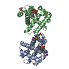
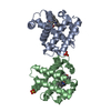
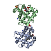
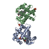
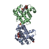
- PDB-3vkm: Protease-resistant mutant form of Human Galectin-8 in complex wit... -
+ Open data

- Basic information
| Entry | Database: PDB / ID: 3vkm | |||||||||
|---|---|---|---|---|---|---|---|---|---|---|
| Title | Protease-resistant mutant form of Human Galectin-8 in complex with sialyllactose and lactose | |||||||||
 Components Components | Galectin-8 | |||||||||
 Keywords Keywords | SUGAR BINDING PROTEIN / BETA-SANDWICH / CARBOHYDRATE BINDING / OLIGOSACCHARIDE | |||||||||
| Function / homology |  Function and homology information Function and homology informationlymphatic endothelial cell migration / xenophagy / plasma cell differentiation / T cell costimulation / cellular response to virus / integrin binding / carbohydrate binding / cytoplasmic vesicle / : / membrane ...lymphatic endothelial cell migration / xenophagy / plasma cell differentiation / T cell costimulation / cellular response to virus / integrin binding / carbohydrate binding / cytoplasmic vesicle / : / membrane / cytosol / cytoplasm Similarity search - Function | |||||||||
| Biological species |  Homo sapiens (human) Homo sapiens (human) | |||||||||
| Method |  X-RAY DIFFRACTION / SYNCHROTRON / MOLECULAR REPLACEMENT / Resolution: 2.98 Å X-RAY DIFFRACTION / SYNCHROTRON / MOLECULAR REPLACEMENT / Resolution: 2.98 Å | |||||||||
 Authors Authors | Yoshida, H. / Yamashita, S. / Teraoka, M. / Nakakita, S. / Nishi, N. / Kamitori, S. | |||||||||
 Citation Citation | Journal: Febs J. / Year: 2012 Title: X-ray structure of a protease-resistant mutant form of human galectin-8 with two carbohydrate recognition domains Authors: Yoshida, H. / Yamashita, S. / Teraoka, M. / Itoh, A. / Nakakita, S. / Nishi, N. / Kamitori, S. | |||||||||
| History |
| |||||||||
| Remark 650 | HELIX Determination method: Author determined | |||||||||
| Remark 700 | SHEET Determination method: Author determined |
- Structure visualization
| Structure viewer | Molecule: MolmilJmol/JSmol |
|---|
- Downloads & links
-Download
| PDBx/mmCIF format | 3vkm.cif.gz | 138 KB | Display | PDBx/mmCIF format |
|---|---|---|---|---|
| PDB format | pdb3vkm.ent.gz | 106.5 KB | Display | PDB format |
| PDBx/mmJSON format | 3vkm.json.gz | Tree view | PDBx/mmJSON format | |
| Others |  Other downloads Other downloads |
-Validation report
| Arichive directory | https://data.pdbj.org/pub/pdb/validation_reports/vk/3vkmftp://data.pdbj.org/pub/pdb/validation_reports/vk/3vkm | HTTPS FTP |
|---|
-Related structure data
| Related structure data |  3vklC  3vknC  3vkoC  4fqzC 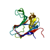 2yv8S C: citing same article ( S: Starting model for refinement |
|---|---|
| Similar structure data |


-Links
 PDBj
PDBj


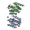
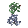
- Assembly
| Deposited unit | 
| ||||||||
|---|---|---|---|---|---|---|---|---|---|
| 1 | 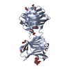
| ||||||||
| 2 | 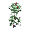
| ||||||||
| Unit cell |
| ||||||||
| Details | AUTHOR STATES THAT THE BIOLOGICAL ASSEMBLY IS MONOMER OR PSEUDO-DIMER OR UNKNOWN. |
-Components
| #1: Protein | Mass: 33110.141 Da / Num. of mol.: 2 Fragment: N-terminal carbohydrate recognition domain, C-terminal carbohydrate recognition domain Source method: isolated from a genetically manipulated source Source: (gene. exp.) Homo sapiens (human) / Gene: LGALS8 / Production host:  #2: Polysaccharide | Source method: isolated from a genetically manipulated source #3: Polysaccharide |   Source method: isolated from a genetically manipulated source Details: oligosaccharide / References: beta-lactose #4: Chemical | ChemComp-EDO /   Mass: 62.068 Da / Num. of mol.: 24 / Source method: obtained synthetically / Formula: C2H6O2 Mass: 62.068 Da / Num. of mol.: 24 / Source method: obtained synthetically / Formula: C2H6O2#5: Water | ChemComp-HOH / |  Mass: 18.015 Da / Num. of mol.: 146 / Source method: isolated from a natural source / Formula: H2O Mass: 18.015 Da / Num. of mol.: 146 / Source method: isolated from a natural source / Formula: H2O |
|---|
-Experimental details
-Experiment
| Experiment | Method: X-RAY DIFFRACTION / Number of used crystals: 1 |
|---|
- Sample preparation
| Crystal | Density Matthews: 2.96 Å3/Da / Density % sol: 58.39 % |
|---|---|
| Crystal grow | Temperature: 293 K / Method: vapor diffusion, hanging drop / pH: 9 Details: 2-3%(v/v) 1,4-dioxane, 9-10%(w/v) PEG20000, 0.1M bicine, pH 9.0, VAPOR DIFFUSION, HANGING DROP, temperature 293K |
-Data collection
| Diffraction | Mean temperature: 100 K |
|---|---|
| Diffraction source | Source: SYNCHROTRON / Site: Photon Factory  / Beamline: AR-NW12A / Wavelength: 1 Å / Beamline: AR-NW12A / Wavelength: 1 Å |
| Detector | Type: ADSC QUANTUM 210r / Detector: CCD / Date: Nov 9, 2010 |
| Radiation | Monochromator: double crystal / Protocol: SINGLE WAVELENGTH / Monochromatic (M) / Laue (L): M / Scattering type: x-ray |
| Radiation wavelength | Wavelength: 1 Å / Relative weight: 1 |
| Reflection | Resolution: 2.98→50 Å / Num. obs: 16883 / % possible obs: 99.7 % / Observed criterion σ(F): 0 / Observed criterion σ(I): 0 / Redundancy: 5.4 % / Biso Wilson estimate: 1.8 Å2 / Rmerge(I) obs: 0.087 / Net I/σ(I): 16.9 |
| Reflection shell | Resolution: 2.98→3.03 Å / Redundancy: 5.1 % / Rmerge(I) obs: 0.497 / Mean I/σ(I) obs: 5.1 / Num. unique all: 822 / % possible all: 100 |
- Processing
| Software |
| |||||||||||||||||||||||||||
|---|---|---|---|---|---|---|---|---|---|---|---|---|---|---|---|---|---|---|---|---|---|---|---|---|---|---|---|---|
| Refinement | Method to determine structure: MOLECULAR REPLACEMENT Starting model: 2YV8 Resolution: 2.98→48.72 Å / Rfactor Rfree error: 0.007 / Occupancy max: 1 / Occupancy min: 0.84 / Data cutoff high absF: 93295 / Data cutoff low absF: 0 / Isotropic thermal model: RESTRAINED / Cross valid method: THROUGHOUT / σ(F): 0 / Stereochemistry target values: Engh & Huber
| |||||||||||||||||||||||||||
| Solvent computation | Solvent model: FLAT MODEL / Bsol: 23.2291 Å2 / ksol: 0.3283 e/Å3 | |||||||||||||||||||||||||||
| Displacement parameters | Biso max: 83.37 Å2 / Biso mean: 49.6472 Å2 / Biso min: 16.11 Å2
| |||||||||||||||||||||||||||
| Refine analyze |
| |||||||||||||||||||||||||||
| Refinement step | Cycle: LAST / Resolution: 2.98→48.72 Å
| |||||||||||||||||||||||||||
| Refine LS restraints |
| |||||||||||||||||||||||||||
| LS refinement shell | Resolution: 2.98→3.17 Å / Rfactor Rfree error: 0.025 / Total num. of bins used: 6
| |||||||||||||||||||||||||||
| Xplor file |
|
