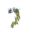
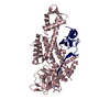
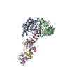
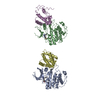
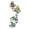
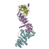
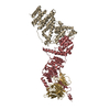
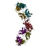
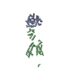
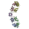
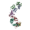
登録情報 データベース : PDB / ID : 6v9iタイトル cryo-EM structure of Cullin5 bound to RING-box protein 2 (Cul5-Rbx2) Immunoglobulin G-binding protein G,Cullin-5 RING-box protein 2 キーワード / / 機能・相同性 分子機能 ドメイン・相同性 構成要素
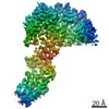
/ / / / / / / / / / / / / / / / / / / / / / / / / / / / / / / / / / / / / / / / / / / / / / / / / / / / / / / / / / / / / / / / / / / / / / / / / / / / / / / / / / / / / / / / / / / / / 生物種 Streptococcus sp. group G (バクテリア)Homo sapiens (ヒト)Mus musculus (ハツカネズミ)手法 / / / 解像度 : 5.2 Å データ登録者 Komives, E.A. / Lumpkin, R.J. / Baker, R.W. / Leschziner, A.E. 資金援助 組織 認可番号 国 National Science Foundation (NSF, United States) 1817774
ジャーナル : Nat Commun / 年 : 2020タイトル : Structure and dynamics of the ASB9 CUL-RING E3 Ligase.著者 : Ryan J Lumpkin / Richard W Baker / Andres E Leschziner / Elizabeth A Komives / 要旨 : The Cullin 5 (CUL5) Ring E3 ligase uses adaptors Elongins B and C (ELOB/C) to bind different SOCS-box-containing substrate receptors, determining the substrate specificity of the ligase. The 18- ... The Cullin 5 (CUL5) Ring E3 ligase uses adaptors Elongins B and C (ELOB/C) to bind different SOCS-box-containing substrate receptors, determining the substrate specificity of the ligase. The 18-member ankyrin and SOCS box (ASB) family is the largest substrate receptor family. Here we report cryo-EM data for the substrate, creatine kinase (CKB) bound to ASB9-ELOB/C, and for full-length CUL5 bound to the RING protein, RBX2, which binds various E2s. To date, no full structures are available either for a substrate-bound ASB nor for CUL5. Hydrogen-deuterium exchange (HDX-MS) mapped onto a full structural model of the ligase revealed long-range allostery extending from the substrate through CUL5. We propose a revised allosteric mechanism for how CUL-E3 ligases function. ASB9 and CUL5 behave as rigid rods, connected through a hinge provided by ELOB/C transmitting long-range allosteric crosstalk from the substrate through CUL5 to the RBX2 flexible linker. 履歴 登録 2019年12月13日 登録サイト / 処理サイト 改定 1.0 2020年4月29日 Provider / タイプ 改定 1.1 2020年11月11日 Group / カテゴリ / citation_authorItem _citation.country / _citation.journal_abbrev ... _citation.country / _citation.journal_abbrev / _citation.journal_id_CSD / _citation.journal_id_ISSN / _citation.journal_volume / _citation.page_first / _citation.page_last / _citation.pdbx_database_id_DOI / _citation.pdbx_database_id_PubMed / _citation.title / _citation.year / _citation_author.identifier_ORCID / _citation_author.name 改定 1.2 2024年10月16日 Group Data collection / Database references ... Data collection / Database references / Refinement description / Structure summary カテゴリ chem_comp_atom / chem_comp_bond ... chem_comp_atom / chem_comp_bond / database_2 / em_3d_fitting_list / em_admin / pdbx_entry_details / pdbx_initial_refinement_model / pdbx_modification_feature Item _database_2.pdbx_DOI / _database_2.pdbx_database_accession ... _database_2.pdbx_DOI / _database_2.pdbx_database_accession / _em_3d_fitting_list.accession_code / _em_3d_fitting_list.initial_refinement_model_id / _em_3d_fitting_list.source_name / _em_3d_fitting_list.type / _em_admin.last_update / _pdbx_entry_details.has_protein_modification
すべて表示 表示を減らす
 ムービー
ムービー コントローラー


 データを開く
 基本情報
基本情報 要素
要素 キーワード
キーワード 機能・相同性情報
 Streptococcus sp. group G (バクテリア)
 Homo sapiens (ヒト)
 データ登録者
 米国, 1件
  引用
引用 構造の表示
構造の表示 ダウンロードとリンク
 その他のダウンロード
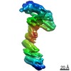

 PDBj
PDBj


 集合体
集合体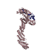

 分子量: 65.409 Da / 分子数: 3 / 由来タイプ: 合成 / 式: Zn
 試料調製
試料調製 電子顕微鏡撮影
 FIELD EMISSION GUN / 加速電圧: 200 kV / 照射モード: FLOOD BEAM
 解析
解析