 Movie
Movie Controller

[English] 日本語
 Yorodumi
- PDB-6uyg: Structure of Hepatitis C Virus Envelope Glycoprotein E2c3 core fr... -
+ Open data

- Basic information
| Entry | Database: PDB / ID: 6uyg | |||||||||||||||||||||
|---|---|---|---|---|---|---|---|---|---|---|---|---|---|---|---|---|---|---|---|---|---|---|
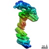
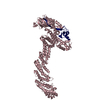
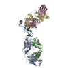
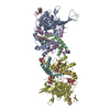
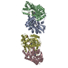
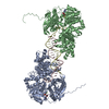
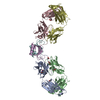
| Title | Structure of Hepatitis C Virus Envelope Glycoprotein E2c3 core from genotype 6a bound to broadly neutralizing antibody AR3A and non neutralizing antibody E1 | |||||||||||||||||||||
 Components Components |
| |||||||||||||||||||||
 Keywords Keywords | IMMUNE SYSTEM / HCV / E2 core / Vaccine design / self-assembly nanoparticle / broadly neutralizing antibodies / bNAbs | |||||||||||||||||||||
| Function / homology |  Function and homology information Function and homology informationIgG binding / host cell mitochondrial membrane / host cell lipid droplet / symbiont-mediated transformation of host cell / symbiont-mediated suppression of host TRAF-mediated signal transduction / symbiont-mediated perturbation of host cell cycle G1/S transition checkpoint / symbiont-mediated suppression of host JAK-STAT cascade via inhibition of STAT1 activity / symbiont-mediated suppression of host cytoplasmic pattern recognition receptor signaling pathway via inhibition of MAVS activity / ribonucleoside triphosphate phosphatase activity / viral nucleocapsid ...IgG binding / host cell mitochondrial membrane / host cell lipid droplet / symbiont-mediated transformation of host cell / symbiont-mediated suppression of host TRAF-mediated signal transduction / symbiont-mediated perturbation of host cell cycle G1/S transition checkpoint / symbiont-mediated suppression of host JAK-STAT cascade via inhibition of STAT1 activity / symbiont-mediated suppression of host cytoplasmic pattern recognition receptor signaling pathway via inhibition of MAVS activity / ribonucleoside triphosphate phosphatase activity / viral nucleocapsid / channel activity / monoatomic ion transmembrane transport / clathrin-dependent endocytosis of virus by host cell / RNA helicase activity / host cell perinuclear region of cytoplasm / host cell endoplasmic reticulum membrane / symbiont-mediated suppression of host type I interferon-mediated signaling pathway / ribonucleoprotein complex / serine-type endopeptidase activity / symbiont-mediated activation of host autophagy / cysteine-type endopeptidase activity / viral RNA genome replication / RNA-directed RNA polymerase activity / fusion of virus membrane with host endosome membrane / viral envelope / virion attachment to host cell / host cell nucleus / host cell plasma membrane / virion membrane / structural molecule activity / proteolysis / RNA binding / zinc ion binding / ATP binding Similarity search - Function | |||||||||||||||||||||
| Biological species |  Recombinant Hepatitis C virus HK6a/JFH-1 Recombinant Hepatitis C virus HK6a/JFH-1 Homo sapiens (human) Homo sapiens (human) Streptococcus sp. (bacteria) Streptococcus sp. (bacteria) | |||||||||||||||||||||
| Method |  X-RAY DIFFRACTION / SYNCHROTRON / MOLECULAR REPLACEMENT / Resolution: 3.375 Å X-RAY DIFFRACTION / SYNCHROTRON / MOLECULAR REPLACEMENT / Resolution: 3.375 Å | |||||||||||||||||||||
 Authors Authors | Tzarum, N. / Wilson, I.A. / Zhu, J. | |||||||||||||||||||||
| Funding support |  United States, 6items United States, 6items
| |||||||||||||||||||||
 Citation Citation | Journal: Sci Adv / Year: 2020 Title: Proof of concept for rational design of hepatitis C virus E2 core nanoparticle vaccines. Authors: He, L. / Tzarum, N. / Lin, X. / Shapero, B. / Sou, C. / Mann, C.J. / Stano, A. / Zhang, L. / Nagy, K. / Giang, E. / Law, M. / Wilson, I.A. / Zhu, J. | |||||||||||||||||||||
| History |
|
- Structure visualization
| Structure viewer | Molecule: MolmilJmol/JSmol |
|---|
- Downloads & links
-Download
| PDBx/mmCIF format | 6uyg.cif.gz | 214.3 KB | Display | PDBx/mmCIF format |
|---|---|---|---|---|
| PDB format | pdb6uyg.ent.gz | 164.7 KB | Display | PDB format |
| PDBx/mmJSON format | 6uyg.json.gz | Tree view | PDBx/mmJSON format | |
| Others |  Other downloads Other downloads |
-Validation report
| Arichive directory | https://data.pdbj.org/pub/pdb/validation_reports/uy/6uygftp://data.pdbj.org/pub/pdb/validation_reports/uy/6uyg | HTTPS FTP |
|---|
-Related structure data
| Related structure data | 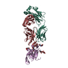 6uydC 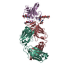 6uyfC  6uymC  6bkbS S: Starting model for refinement C: citing same article ( |
|---|---|
| Similar structure data |

-Links
 PDBj
PDBj


- Assembly
| Deposited unit | 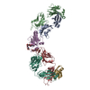
| ||||||||
|---|---|---|---|---|---|---|---|---|---|
| 1 |
| ||||||||
| Unit cell |
|
-Components
-Protein , 1 types, 1 molecules E
| #1: Protein | Mass: 20707.395 Da / Num. of mol.: 1 Source method: isolated from a genetically manipulated source Source: (gene. exp.) Recombinant Hepatitis C virus HK6a/JFH-1Production host: Homo sapiens (human) / References: UniProt: B9V0E2*PLUS |
|---|
-Antibody , 5 types, 5 molecules HLABG
| #2: Antibody | Mass: 24581.408 Da / Num. of mol.: 1 Source method: isolated from a genetically manipulated source Source: (gene. exp.) Homo sapiens (human) / Production host: Homo sapiens (human) |
|---|---|
| #3: Antibody | Mass: 23296.824 Da / Num. of mol.: 1 Source method: isolated from a genetically manipulated source Source: (gene. exp.) Homo sapiens (human) / Production host: Homo sapiens (human) |
| #4: Antibody | Mass: 23937.957 Da / Num. of mol.: 1 Source method: isolated from a genetically manipulated source Source: (gene. exp.) Homo sapiens (human) / Production host: Homo sapiens (human) |
| #5: Antibody | Mass: 24051.750 Da / Num. of mol.: 1 Source method: isolated from a genetically manipulated source Source: (gene. exp.) Homo sapiens (human) / Production host: Homo sapiens (human) |
| #6: Antibody | Mass: 6657.354 Da / Num. of mol.: 1 Source method: isolated from a genetically manipulated source Source: (gene. exp.) Streptococcus sp. (bacteria) / Production host: |
-Sugars , 2 types, 3 molecules 
| #7: Polysaccharide | 2-acetamido-2-deoxy-beta-D-glucopyranose-(1-4)-2-acetamido-2-deoxy-beta-D-glucopyranose Source method: isolated from a genetically manipulated source |
|---|---|
| #8: Sugar |  Type: D-saccharide, beta linking / Mass: 221.208 Da / Num. of mol.: 2 Type: D-saccharide, beta linking / Mass: 221.208 Da / Num. of mol.: 2Source method: isolated from a genetically manipulated source Formula: C8H15NO6 / Feature type: SUBJECT OF INVESTIGATION |
-Details
| Has ligand of interest | Y |
|---|---|
| Has protein modification | Y |
-Experimental details
-Experiment
| Experiment | Method: X-RAY DIFFRACTION / Number of used crystals: 1 |
|---|
- Sample preparation
| Crystal | Density Matthews: 3.61 Å3/Da / Density % sol: 65.91 % |
|---|---|
| Crystal grow | Temperature: 293 K / Method: vapor diffusion, sitting drop Details: 0.2M magnesium chloride, 10% (w/v) PEG 3000, 15% ethylene glycol, 0.1M Na-cacodylate, pH=6.5 |
-Data collection
| Diffraction | Mean temperature: 100 K / Serial crystal experiment: N |
|---|---|
| Diffraction source | Source: SYNCHROTRON / Site: SSRL / Beamline: BL12-2 / Wavelength: 0.97946 Å |
| Detector | Type: DECTRIS PILATUS3 S 6M / Detector: PIXEL / Date: Jan 25, 2018 |
| Radiation | Protocol: SINGLE WAVELENGTH / Monochromatic (M) / Laue (L): M / Scattering type: x-ray |
| Radiation wavelength | Wavelength: 0.97946 Å / Relative weight: 1 |
| Reflection | Resolution: 3.375→30 Å / Num. obs: 25916 / % possible obs: 100 % / Redundancy: 17.2 % / CC1/2: 0.92 / Net I/σ(I): 13.5 |
| Reflection shell | Resolution: 3.4→3.46 Å / Num. unique obs: 1258 / CC1/2: 0.29 |
- Processing
| Software |
| ||||||||||||||||||||||||||||||||||||||||||||||||||||||||||||
|---|---|---|---|---|---|---|---|---|---|---|---|---|---|---|---|---|---|---|---|---|---|---|---|---|---|---|---|---|---|---|---|---|---|---|---|---|---|---|---|---|---|---|---|---|---|---|---|---|---|---|---|---|---|---|---|---|---|---|---|---|---|
| Refinement | Method to determine structure: MOLECULAR REPLACEMENT Starting model: 6BKB Resolution: 3.375→29.6 Å / SU ML: 0.46 / Cross valid method: THROUGHOUT / σ(F): 1.35 / Phase error: 33.31
| ||||||||||||||||||||||||||||||||||||||||||||||||||||||||||||
| Solvent computation | Shrinkage radii: 0.9 Å / VDW probe radii: 1.11 Å | ||||||||||||||||||||||||||||||||||||||||||||||||||||||||||||
| Displacement parameters | Biso max: 230.33 Å2 / Biso mean: 102.8073 Å2 / Biso min: 30 Å2 | ||||||||||||||||||||||||||||||||||||||||||||||||||||||||||||
| Refinement step | Cycle: final / Resolution: 3.375→29.6 Å
| ||||||||||||||||||||||||||||||||||||||||||||||||||||||||||||
| Refine LS restraints |
| ||||||||||||||||||||||||||||||||||||||||||||||||||||||||||||
| LS refinement shell | Refine-ID: X-RAY DIFFRACTION / Rfactor Rfree error: 0
|
