 Movie
Movie Controller

[English] 日本語
 Yorodumi
- PDB-1ung: Structural mechanism for the inhibition of CDK5-p25 by roscovitin... -
+ Open data

- Basic information
| Entry | Database: PDB / ID: 1ung | ||||||
|---|---|---|---|---|---|---|---|
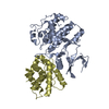
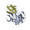
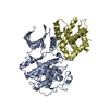
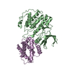
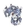
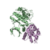
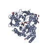
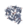
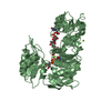
| Title | Structural mechanism for the inhibition of CDK5-p25 by roscovitine, aloisine and indirubin. | ||||||
 Components Components |
| ||||||
 Keywords Keywords | CELL CYCLE / COMPLEX(KINASE-ACTIVATOR) / INHIBITORS / NEURODEGENERATIVE DISEASES | ||||||
| Function / homology |  Function and homology information Function and homology informationpositive regulation of presynaptic cytosolic calcium concentration / negative regulation of calcium ion-dependent exocytosis of neurotransmitter / superior olivary nucleus maturation / acetylcholine receptor activator activity / protein kinase 5 complex / cerebellar cortex formation / G1 to G0 transition involved in cell differentiation / ErbB-2 class receptor binding / negative regulation of synaptic plasticity / regulation of cell cycle phase transition ...positive regulation of presynaptic cytosolic calcium concentration / negative regulation of calcium ion-dependent exocytosis of neurotransmitter / superior olivary nucleus maturation / acetylcholine receptor activator activity / protein kinase 5 complex / cerebellar cortex formation / G1 to G0 transition involved in cell differentiation / ErbB-2 class receptor binding / negative regulation of synaptic plasticity / regulation of cell cycle phase transition / contractile muscle fiber / Activated NTRK2 signals through CDK5 / layer formation in cerebral cortex / corpus callosum development / positive regulation of calcium ion-dependent exocytosis / negative regulation of axon extension / neuron cell-cell adhesion / receptor catabolic process / protein localization to synapse / NGF-stimulated transcription / CRMPs in Sema3A signaling / calcium ion import / axon extension / regulation of dendritic spine morphogenesis / signaling receptor inhibitor activity / ErbB-3 class receptor binding / negative regulation of protein export from nucleus / axonal fasciculation / regulation of cyclin-dependent protein serine/threonine kinase activity / dendrite morphogenesis / cyclin-dependent protein serine/threonine kinase activator activity / Phosphorylation and nuclear translocation of BMAL1 (ARNTL) and CLOCK / motor neuron axon guidance / regulation of neuron differentiation / regulation of synaptic vesicle recycling / Phosphorylation and nuclear translocation of the CRY:PER:kinase complex / tau-protein kinase activity / Deregulated CDK5 triggers multiple neurodegenerative pathways in Alzheimer's disease models / peptidyl-threonine phosphorylation / central nervous system neuron development / oligodendrocyte differentiation / receptor clustering / negative regulation of cell cycle / synaptic vesicle transport / synaptic transmission, dopaminergic / synaptic vesicle exocytosis / ephrin receptor signaling pathway / DARPP-32 events / regulation of macroautophagy / positive regulation of protein targeting to membrane / protein kinase activator activity / cyclin-dependent protein serine/threonine kinase activity / Schwann cell development / skeletal muscle tissue development / synaptic vesicle endocytosis / alpha-tubulin binding / regulation of protein localization to plasma membrane / beta-tubulin binding / regulation of synaptic transmission, glutamatergic / cyclin-dependent protein kinase holoenzyme complex / positive regulation of microtubule polymerization / NPAS4 regulates expression of target genes / negative regulation of proteolysis / behavioral response to cocaine / axonogenesis / synapse assembly / negative regulation of protein ubiquitination / ionotropic glutamate receptor signaling pathway / regulation of cell migration / ionotropic glutamate receptor binding / sensory perception of pain / cerebellum development / axon guidance / excitatory postsynaptic potential / cell-matrix adhesion / protein serine/threonine kinase activator activity / peptidyl-serine phosphorylation / hippocampus development / regulation of actin cytoskeleton organization / filopodium / neuromuscular junction / synaptic transmission, glutamatergic / brain development / visual learning / Hsp90 protein binding / intracellular protein transport / regulation of synaptic plasticity / microtubule cytoskeleton organization / neuron migration / tau protein binding / cellular response to amyloid-beta / neuron projection development / neuron differentiation / cell junction / kinase activity / p53 binding / actin filament binding / G protein-coupled acetylcholine receptor signaling pathway / rhythmic process / positive regulation of neuron apoptotic process Similarity search - Function | ||||||
| Biological species |  HOMO SAPIENS (human) HOMO SAPIENS (human) | ||||||
| Method |  X-RAY DIFFRACTION / SYNCHROTRON / MOLECULAR REPLACEMENT / Resolution: 2.3 Å X-RAY DIFFRACTION / SYNCHROTRON / MOLECULAR REPLACEMENT / Resolution: 2.3 Å | ||||||
 Authors Authors | Mapelli, M. / Crovace, C. / Massimiliano, L. / Musacchio, A. | ||||||
 Citation Citation | Journal: J.Med.Chem. / Year: 2005 Title: Mechanism of Cdk5/P25 Binding by Cdk Inhibitors Authors: Mapelli, M. / Massimilinao, L. / Crovace, C. / Seeliger, M.A. / Tsai, L.-H. / Meijer, L. / Musacchio, A. | ||||||
| History |
|
- Structure visualization
| Structure viewer | Molecule: MolmilJmol/JSmol |
|---|
- Downloads & links
-Download
| PDBx/mmCIF format | 1ung.cif.gz | 188.1 KB | Display | PDBx/mmCIF format |
|---|---|---|---|---|
| PDB format | pdb1ung.ent.gz | 150.1 KB | Display | PDB format |
| PDBx/mmJSON format | 1ung.json.gz | Tree view | PDBx/mmJSON format | |
| Others |  Other downloads Other downloads |
-Validation report
| Arichive directory | https://data.pdbj.org/pub/pdb/validation_reports/un/1ungftp://data.pdbj.org/pub/pdb/validation_reports/un/1ung | HTTPS FTP |
|---|
-Related structure data
| Related structure data |  1unhC  1unlC  1h4lS S: Starting model for refinement C: citing same article ( |
|---|---|
| Similar structure data |


-Links
 PDBj
PDBj


- Assembly
| Deposited unit | 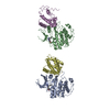
| ||||||||
|---|---|---|---|---|---|---|---|---|---|
| 1 | 
| ||||||||
| 2 | 
| ||||||||
| Unit cell |
|
-Components
| #1: Protein | Mass: 33349.477 Da / Num. of mol.: 2 / Mutation: YES Source method: isolated from a genetically manipulated source Source: (gene. exp.) HOMO SAPIENS (human) / Plasmid: PFASTBAC / Cell line (production host): SF9 / Production host:   SPODOPTERA FRUGIPERDA (fall armyworm) / References: UniProt: Q00535, cyclin-dependent kinase SPODOPTERA FRUGIPERDA (fall armyworm) / References: UniProt: Q00535, cyclin-dependent kinase#2: Protein | Mass: 23200.678 Da / Num. of mol.: 2 / Fragment: RESIDUES 100-307 Source method: isolated from a genetically manipulated source Source: (gene. exp.) HOMO SAPIENS (human) / Plasmid: PFASTBAC / Cell line (production host): SF9 / Production host: SPODOPTERA FRUGIPERDA (fall armyworm) / References: UniProt: Q15078#3: Chemical |   Mass: 267.326 Da / Num. of mol.: 2 / Source method: obtained synthetically / Formula: C16H17N3O Mass: 267.326 Da / Num. of mol.: 2 / Source method: obtained synthetically / Formula: C16H17N3O#4: Water | ChemComp-HOH / |  Mass: 18.015 Da / Num. of mol.: 294 / Source method: isolated from a natural source / Formula: H2O Mass: 18.015 Da / Num. of mol.: 294 / Source method: isolated from a natural source / Formula: H2OCompound details | ENGINEERED | |
|---|
-Experimental details
-Experiment
| Experiment | Method: X-RAY DIFFRACTION / Number of used crystals: 1 |
|---|
- Sample preparation
| Crystal | Density Matthews: 2.8 Å3/Da / Density % sol: 56.4 % |
|---|---|
| Crystal grow | pH: 7 Details: 13% PEG 3350, 0.1M KI, 0.1M BISTRISPROPANE PH 7.0, 10MM DTT |
-Data collection
| Diffraction | Mean temperature: 287 K |
|---|---|
| Diffraction source | Source: SYNCHROTRON / Site: ESRF  / Beamline: ID14-2 / Wavelength: 0.933 / Beamline: ID14-2 / Wavelength: 0.933 |
| Detector | Type: ADSC CCD / Detector: CCD / Date: Sep 15, 2002 |
| Radiation | Protocol: SINGLE WAVELENGTH / Monochromatic (M) / Laue (L): M / Scattering type: x-ray |
| Radiation wavelength | Wavelength: 0.933 Å / Relative weight: 1 |
| Reflection | Resolution: 2.3→25 Å / Num. obs: 53731 / % possible obs: 95.4 % / Observed criterion σ(I): 2.5 / Redundancy: 6.2 % / Rmerge(I) obs: 0.104 / Net I/σ(I): 9.1 |
| Reflection shell | Resolution: 2.3→2.4 Å / Redundancy: 2.8 % / Rmerge(I) obs: 0.37 / Mean I/σ(I) obs: 2.6 / % possible all: 97.5 |
- Processing
| Software |
| ||||||||||||||||||||||||||||||||||||||||||||||||||||||||||||||||||||||||||||||||||||||||||||||||||||||||||||||||||||||||||||||||||||||||||||||||||||||||||||||||||||||||||||||||||||||
|---|---|---|---|---|---|---|---|---|---|---|---|---|---|---|---|---|---|---|---|---|---|---|---|---|---|---|---|---|---|---|---|---|---|---|---|---|---|---|---|---|---|---|---|---|---|---|---|---|---|---|---|---|---|---|---|---|---|---|---|---|---|---|---|---|---|---|---|---|---|---|---|---|---|---|---|---|---|---|---|---|---|---|---|---|---|---|---|---|---|---|---|---|---|---|---|---|---|---|---|---|---|---|---|---|---|---|---|---|---|---|---|---|---|---|---|---|---|---|---|---|---|---|---|---|---|---|---|---|---|---|---|---|---|---|---|---|---|---|---|---|---|---|---|---|---|---|---|---|---|---|---|---|---|---|---|---|---|---|---|---|---|---|---|---|---|---|---|---|---|---|---|---|---|---|---|---|---|---|---|---|---|---|---|
| Refinement | Method to determine structure: MOLECULAR REPLACEMENT Starting model: PDB ENTRY 1H4L Resolution: 2.3→19.76 Å / Cor.coef. Fo:Fc: 0.923 / Cor.coef. Fo:Fc free: 0.921 / TLS residual ADP flag: LIKELY RESIDUAL / Cross valid method: THROUGHOUT / ESU R: 0.293 / ESU R Free: 0.201 / Stereochemistry target values: MAXIMUM LIKELIHOOD / Details: HYDROGENS HAVE BEEN ADDED IN THE RIDING POSITIONS
| ||||||||||||||||||||||||||||||||||||||||||||||||||||||||||||||||||||||||||||||||||||||||||||||||||||||||||||||||||||||||||||||||||||||||||||||||||||||||||||||||||||||||||||||||||||||
| Solvent computation | Ion probe radii: 0.8 Å / Shrinkage radii: 0.8 Å / VDW probe radii: 1.4 Å / Solvent model: BABINET MODEL WITH MASK | ||||||||||||||||||||||||||||||||||||||||||||||||||||||||||||||||||||||||||||||||||||||||||||||||||||||||||||||||||||||||||||||||||||||||||||||||||||||||||||||||||||||||||||||||||||||
| Displacement parameters | Biso mean: 22.42 Å2
| ||||||||||||||||||||||||||||||||||||||||||||||||||||||||||||||||||||||||||||||||||||||||||||||||||||||||||||||||||||||||||||||||||||||||||||||||||||||||||||||||||||||||||||||||||||||
| Refinement step | Cycle: LAST / Resolution: 2.3→19.76 Å
| ||||||||||||||||||||||||||||||||||||||||||||||||||||||||||||||||||||||||||||||||||||||||||||||||||||||||||||||||||||||||||||||||||||||||||||||||||||||||||||||||||||||||||||||||||||||
| Refine LS restraints |
|