Movie
Movie Controller

[English] 日本語
 Yorodumi
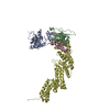
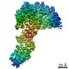
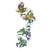
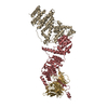
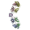
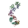
- PDB-6v9i: cryo-EM structure of Cullin5 bound to RING-box protein 2 (Cul5-Rbx2) -
+ Open data

- Basic information
| Entry | Database: PDB / ID: 6v9i | ||||||
|---|---|---|---|---|---|---|---|
| Title | cryo-EM structure of Cullin5 bound to RING-box protein 2 (Cul5-Rbx2) | ||||||
 Components Components |
| ||||||
 Keywords Keywords | LIGASE / E3 Ubiquitin Ligase / RING-box protein | ||||||
| Function / homology |  Function and homology information Function and homology informationradial glia guided migration of Purkinje cell / negative regulation of growth hormone receptor signaling pathway / negative regulation of focal adhesion disassembly / Inactivation of CSF3 (G-CSF) signaling / phosphocreatine biosynthetic process / cerebral cortex radially oriented cell migration / ERBB2 signaling pathway / negative regulation of focal adhesion assembly / reelin-mediated signaling pathway / Neddylation ...radial glia guided migration of Purkinje cell / negative regulation of growth hormone receptor signaling pathway / negative regulation of focal adhesion disassembly / Inactivation of CSF3 (G-CSF) signaling / phosphocreatine biosynthetic process / cerebral cortex radially oriented cell migration / ERBB2 signaling pathway / negative regulation of focal adhesion assembly / reelin-mediated signaling pathway / Neddylation / erythropoietin-mediated signaling pathway / cullin-RING-type E3 NEDD8 transferase / regulation of neuron migration / Antigen processing: Ubiquitination & Proteasome degradation / protein K11-linked ubiquitination / protein neddylation / NEDD8 ligase activity / IgG binding / cGAS/STING signaling pathway / growth hormone receptor signaling pathway / intrinsic apoptotic signaling pathway in response to oxidative stress / Cul5-RING ubiquitin ligase complex / SCF ubiquitin ligase complex / SCF-dependent proteasomal ubiquitin-dependent protein catabolic process / apoptotic mitochondrial changes / ubiquitin ligase complex scaffold activity / growth hormone receptor signaling pathway via JAK-STAT / epithelial to mesenchymal transition / positive regulation of focal adhesion assembly / cullin family protein binding / site of DNA damage / endoplasmic reticulum unfolded protein response / proteasomal protein catabolic process / intrinsic apoptotic signaling pathway / G1/S transition of mitotic cell cycle / RING-type E3 ubiquitin transferase / Inactivation of CSF3 (G-CSF) signaling / Vif-mediated degradation of APOBEC3G / Evasion by RSV of host interferon responses / Downregulation of ERBB2 signaling / calcium channel activity / ubiquitin-protein transferase activity / ubiquitin protein ligase activity / cell migration / Antigen processing: Ubiquitination & Proteasome degradation / signaling receptor activity / Neddylation / defense response to virus / ubiquitin-dependent protein catabolic process / proteasome-mediated ubiquitin-dependent protein catabolic process / protein-macromolecule adaptor activity / positive regulation of cell migration / protein ubiquitination / innate immune response / ubiquitin protein ligase binding / negative regulation of apoptotic process / zinc ion binding / nucleoplasm / nucleus / cytoplasm / cytosol Similarity search - Function | ||||||
| Biological species |  Streptococcus sp. group G (bacteria) Streptococcus sp. group G (bacteria) Homo sapiens (human) Homo sapiens (human) | ||||||
| Method | ELECTRON MICROSCOPY / single particle reconstruction / cryo EM / Resolution: 5.2 Å | ||||||
 Authors Authors | Komives, E.A. / Lumpkin, R.J. / Baker, R.W. / Leschziner, A.E. | ||||||
| Funding support |  United States, 1items United States, 1items
| ||||||
 Citation Citation | Journal: Nat Commun / Year: 2020 Title: Structure and dynamics of the ASB9 CUL-RING E3 Ligase. Authors: Ryan J Lumpkin / Richard W Baker / Andres E Leschziner / Elizabeth A Komives / Abstract: The Cullin 5 (CUL5) Ring E3 ligase uses adaptors Elongins B and C (ELOB/C) to bind different SOCS-box-containing substrate receptors, determining the substrate specificity of the ligase. The 18- ...The Cullin 5 (CUL5) Ring E3 ligase uses adaptors Elongins B and C (ELOB/C) to bind different SOCS-box-containing substrate receptors, determining the substrate specificity of the ligase. The 18-member ankyrin and SOCS box (ASB) family is the largest substrate receptor family. Here we report cryo-EM data for the substrate, creatine kinase (CKB) bound to ASB9-ELOB/C, and for full-length CUL5 bound to the RING protein, RBX2, which binds various E2s. To date, no full structures are available either for a substrate-bound ASB nor for CUL5. Hydrogen-deuterium exchange (HDX-MS) mapped onto a full structural model of the ligase revealed long-range allostery extending from the substrate through CUL5. We propose a revised allosteric mechanism for how CUL-E3 ligases function. ASB9 and CUL5 behave as rigid rods, connected through a hinge provided by ELOB/C transmitting long-range allosteric crosstalk from the substrate through CUL5 to the RBX2 flexible linker. | ||||||
| History |
|
- Structure visualization
| Movie |
Movie viewer |
|---|---|
| Structure viewer | Molecule: MolmilJmol/JSmol |
- Downloads & links
-Download
| PDBx/mmCIF format | 6v9i.cif.gz | 132.9 KB | Display | PDBx/mmCIF format |
|---|---|---|---|---|
| PDB format | pdb6v9i.ent.gz | 79.2 KB | Display | PDB format |
| PDBx/mmJSON format | 6v9i.json.gz | Tree view | PDBx/mmJSON format | |
| Others |  Other downloads Other downloads |
-Validation report
| Arichive directory | https://data.pdbj.org/pub/pdb/validation_reports/v9/6v9iftp://data.pdbj.org/pub/pdb/validation_reports/v9/6v9i | HTTPS FTP |
|---|
-Related structure data
| Related structure data | 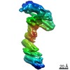 21121MC  6v9hC C: citing same article ( M: map data used to model this data |
|---|---|
| Similar structure data |

-Links
 PDBj
PDBj


- Assembly
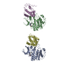
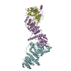
| Deposited unit | 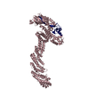
|
|---|---|
| 1 |
|
-Components
| #1: Antibody | Mass: 100456.500 Da / Num. of mol.: 1 Source method: isolated from a genetically manipulated source Source: (gene. exp.) Streptococcus sp. group G (bacteria), (gene. exp.) Homo sapiens (human)Gene: spg, CUL5, VACM1 / Production host: | ||||
|---|---|---|---|---|---|
| #2: Protein | Mass: 12721.500 Da / Num. of mol.: 1 Source method: isolated from a genetically manipulated source Source: (gene. exp.) | ||||
| #3: Chemical |   Mass: 65.409 Da / Num. of mol.: 3 / Source method: obtained synthetically / Formula: Zn Mass: 65.409 Da / Num. of mol.: 3 / Source method: obtained synthetically / Formula: ZnHas ligand of interest | N | Has protein modification | Y | |
-Experimental details
-Experiment
| Experiment | Method: ELECTRON MICROSCOPY |
|---|---|
| EM experiment | Aggregation state: PARTICLE / 3D reconstruction method: single particle reconstruction |
- Sample preparation
| Component | Name: cryo-EM structure of Cullin5 bound to RING-box protein 2 (Cul5-Rbx2) Type: COMPLEX / Entity ID: #1-#2 / Source: RECOMBINANT |
|---|---|
| Molecular weight | Value: 0.1 MDa / Experimental value: NO |
| Source (natural) | Organism: Homo sapiens (human) |
| Source (recombinant) | Organism: |
| Buffer solution | pH: 7.5 |
| Specimen | Conc.: 0.5 mg/ml / Embedding applied: NO / Shadowing applied: NO / Staining applied: NO / Vitrification applied: YES |
| Specimen support | Grid material: GOLD / Grid mesh size: 300 divisions/in. / Grid type: UltrAuFoil |
| Vitrification | Instrument: FEI VITROBOT MARK IV / Cryogen name: ETHANE / Humidity: 100 % / Chamber temperature: 277 K / Details: 4 second blot time, blot force 20 |
- Electron microscopy imaging
| Experimental equipment |  Model: Talos Arctica / Image courtesy: FEI Company |
|---|---|
| Microscopy | Model: FEI TALOS ARCTICA |
| Electron gun | Electron source:  FIELD EMISSION GUN / Accelerating voltage: 200 kV / Illumination mode: FLOOD BEAM FIELD EMISSION GUN / Accelerating voltage: 200 kV / Illumination mode: FLOOD BEAM |
| Electron lens | Mode: BRIGHT FIELD / Nominal magnification: 36000 X / Nominal defocus max: 2000 nm / Nominal defocus min: 1200 nm / Cs: 2.7 mm / C2 aperture diameter: 70 µm / Alignment procedure: COMA FREE |
| Specimen holder | Cryogen: NITROGEN |
| Image recording | Average exposure time: 8 sec. / Electron dose: 59 e/Å2 / Detector mode: COUNTING / Film or detector model: GATAN K2 SUMMIT (4k x 4k) |
- Processing
| EM software |
| ||||||||||||||||||||||||
|---|---|---|---|---|---|---|---|---|---|---|---|---|---|---|---|---|---|---|---|---|---|---|---|---|---|
| CTF correction | Type: PHASE FLIPPING AND AMPLITUDE CORRECTION | ||||||||||||||||||||||||
| Symmetry | Point symmetry: C1 (asymmetric) | ||||||||||||||||||||||||
| 3D reconstruction | Resolution: 5.2 Å / Resolution method: FSC 0.143 CUT-OFF / Num. of particles: 46877 / Details: Non-uniform refinement in cryoSPARC v2 / Symmetry type: POINT | ||||||||||||||||||||||||
| Atomic model building | Protocol: FLEXIBLE FIT / Space: REAL | ||||||||||||||||||||||||
| Atomic model building |
|