National Institutes of Health/National Institute of General Medical Sciences (NIH/NIGMS)
GM43949
United States
Citation
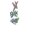
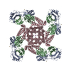
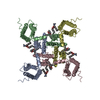
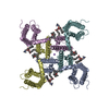
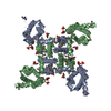
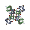
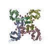
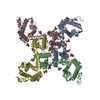
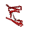
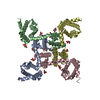
Journal: Elife / Year: 2019 Title: Cryo-EM structure of the KvAP channel reveals a non-domain-swapped voltage sensor topology. Authors: Xiao Tao / Roderick MacKinnon / Abstract: Conductance in voltage-gated ion channels is regulated by membrane voltage through structural domains known as voltage sensors. A single structural class of voltage sensor domain exists, but two ...Conductance in voltage-gated ion channels is regulated by membrane voltage through structural domains known as voltage sensors. A single structural class of voltage sensor domain exists, but two different modes of voltage sensor attachment to the pore occur in nature: domain-swapped and non-domain-swapped. Since the more thoroughly studied Kv1-7, Nav and Cav channels have domain-swapped voltage sensors, much less is known about non-domain-swapped voltage-gated ion channels. In this paper, using cryo-EM, we show that KvAP from has non-domain-swapped voltage sensors as well as other unusual features. The new structure, together with previous functional data, suggests that KvAP and the Shaker channel, to which KvAP is most often compared, probably undergo rather different voltage-dependent conformational changes when they open.
Conc.: 6 mg/ml / Embedding applied: NO / Shadowing applied: NO / Staining applied: NO / Vitrification applied: YES / Details: KvAP in complex with 6E1 Fab
Average exposure time: 10 sec. / Electron dose: 75 e/Å2 / Detector mode: SUPER-RESOLUTION / Film or detector model: GATAN K2 SUMMIT (4k x 4k) / Num. of grids imaged: 2 / Num. of real images: 8000
Image scans
Movie frames/image: 50 / Used frames/image: 1-50
-
Processing
EM software
ID
Name
Version
Category
1
Gautomatch
v0.56_sm61_cu8.0
particleselection
2
SerialEM
imageacquisition
4
Gctf
1.06
CTFcorrection
7
UCSF Chimera
1.13.1
modelfitting
8
Coot
0.8.9.2
modelfitting
10
PHENIX
1.16
modelrefinement
11
cryoSPARC
2.9.0
initialEulerassignment
12
FREALIGN
9.11
finalEulerassignment
13
RELION
3.0.8
classification
14
FREALIGN
9.11
3Dreconstruction
CTF correction
Type: NONE
Particle selection
Num. of particles selected: 1800000
Symmetry
Point symmetry: C4 (4 fold cyclic)
3D reconstruction
Resolution: 5.9 Å / Resolution method: FSC 0.143 CUT-OFF / Num. of particles: 67000 / Num. of class averages: 2 / Symmetry type: POINT
In the structure databanks used in Yorodumi, some data are registered as the other names, "COVID-19 virus" and "2019-nCoV". Here are the details of the virus and the list of structure data.
Jan 31, 2019. EMDB accession codes are about to change! (news from PDBe EMDB page)
EMDB accession codes are about to change! (news from PDBe EMDB page)
The allocation of 4 digits for EMDB accession codes will soon come to an end. Whilst these codes will remain in use, new EMDB accession codes will include an additional digit and will expand incrementally as the available range of codes is exhausted. The current 4-digit format prefixed with “EMD-” (i.e. EMD-XXXX) will advance to a 5-digit format (i.e. EMD-XXXXX), and so on. It is currently estimated that the 4-digit codes will be depleted around Spring 2019, at which point the 5-digit format will come into force.
The EM Navigator/Yorodumi systems omit the EMD- prefix.
Related info.:Q: What is EMD? / ID/Accession-code notation in Yorodumi/EM Navigator
Yorodumi is a browser for structure data from EMDB, PDB, SASBDB, etc.
This page is also the successor to EM Navigator detail page, and also detail information page/front-end page for Omokage search.
The word "yorodu" (or yorozu) is an old Japanese word meaning "ten thousand". "mi" (miru) is to see.
Related info.:EMDB / PDB / SASBDB / Comparison of 3 databanks / Yorodumi Search / Aug 31, 2016. New EM Navigator & Yorodumi / Yorodumi Papers / Jmol/JSmol / Function and homology information / Changes in new EM Navigator and Yorodumi
 Movie
Movie Controller


 Open data
 Basic information
 Components
 Keywords
 Function and homology information
 Aeropyrum pernix (archaea)
 Authors
 United States, 1items
  Citation
 Structure visualization
 Downloads & links
 Other downloads
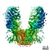
 PDBj
PDBj
 Assembly
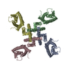

 Sample preparation
 Electron microscopy imaging
 FIELD EMISSION GUN / Accelerating voltage: 300 kV / Illumination mode: FLOOD BEAM
 Processing