 Movie
Movie Controller

[English] 日本語
 Yorodumi
- PDB-5yub: Crystal structure of voltage-gated sodium channel NavAb E32Q mutant -
+ Open data

- Basic information
| Entry | Database: PDB / ID: 5yub | ||||||
|---|---|---|---|---|---|---|---|
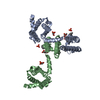
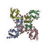
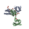
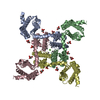
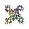
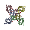
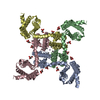
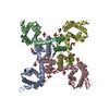
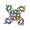
| Title | Crystal structure of voltage-gated sodium channel NavAb E32Q mutant | ||||||
 Components Components | Ion transport protein | ||||||
 Keywords Keywords | MEMBRANE PROTEIN / ion channel | ||||||
| Function / homology |  Function and homology information Function and homology informationvoltage-gated sodium channel complex / voltage-gated sodium channel activity / metal ion binding / identical protein binding Similarity search - Function | ||||||
| Biological species |  Arcobacter butzleri (bacteria) Arcobacter butzleri (bacteria) | ||||||
| Method |  X-RAY DIFFRACTION / SYNCHROTRON / MOLECULAR REPLACEMENT / Resolution: 3.40003993335 Å X-RAY DIFFRACTION / SYNCHROTRON / MOLECULAR REPLACEMENT / Resolution: 3.40003993335 Å | ||||||
 Authors Authors | Irie, K. / Shimomura, T. / Fujiyoshi, Y. | ||||||
| Funding support |  Japan, 1items Japan, 1items
| ||||||
 Citation Citation | Journal: FEBS Lett. / Year: 2017 Title: Structural insight on the voltage dependence of prokaryotic voltage gated sodium channel NavAb. Authors: Irie, K. / Haga, Y. / Shimomura, T. / Fujiyoshi, Y. | ||||||
| History |
|
- Structure visualization
| Structure viewer | Molecule: MolmilJmol/JSmol |
|---|
- Downloads & links
-Download
| PDBx/mmCIF format | 5yub.cif.gz | 125.4 KB | Display | PDBx/mmCIF format |
|---|---|---|---|---|
| PDB format | pdb5yub.ent.gz | 85.9 KB | Display | PDB format |
| PDBx/mmJSON format | 5yub.json.gz | Tree view | PDBx/mmJSON format | |
| Others |  Other downloads Other downloads |
-Validation report
| Arichive directory | https://data.pdbj.org/pub/pdb/validation_reports/yu/5yubftp://data.pdbj.org/pub/pdb/validation_reports/yu/5yub | HTTPS FTP |
|---|
-Related structure data
| Related structure data | 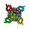 5yuaSC 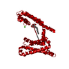 5yucC S: Starting model for refinement C: citing same article ( |
|---|---|
| Similar structure data |

-Links
 PDBj
PDBj
- Assembly
| Deposited unit | 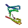
| ||||||||||||
|---|---|---|---|---|---|---|---|---|---|---|---|---|---|
| 1 | 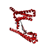
| ||||||||||||
| Unit cell |
| ||||||||||||
| Components on special symmetry positions |
|
-Components
| #1: Protein | Mass: 31381.221 Da / Num. of mol.: 1 / Mutation: E32Q Source method: isolated from a genetically manipulated source Source: (gene. exp.) Arcobacter butzleri (bacteria) / Gene: SAMEA4475700_01771 / Production host: |
|---|---|
| #2: Chemical | ChemComp-CA /   Mass: 40.078 Da / Num. of mol.: 1 / Source method: obtained synthetically / Formula: Ca Mass: 40.078 Da / Num. of mol.: 1 / Source method: obtained synthetically / Formula: Ca |
| #3: Sugar | ChemComp-LMT /   Type: D-saccharide / Mass: 510.615 Da / Num. of mol.: 1 / Source method: obtained synthetically / Formula: C24H46O11 / Comment: detergent*YM Type: D-saccharide / Mass: 510.615 Da / Num. of mol.: 1 / Source method: obtained synthetically / Formula: C24H46O11 / Comment: detergent*YM |
| #4: Chemical | ChemComp-1N7 /   Mass: 631.884 Da / Num. of mol.: 1 / Source method: obtained synthetically / Formula: C32H59N2O8S / Comment: detergent*YM Mass: 631.884 Da / Num. of mol.: 1 / Source method: obtained synthetically / Formula: C32H59N2O8S / Comment: detergent*YM |
-Experimental details
-Experiment
| Experiment | Method: X-RAY DIFFRACTION / Number of used crystals: 1 |
|---|
- Sample preparation
| Crystal | Density Matthews: 6.51 Å3/Da / Density % sol: 81.12 % |
|---|---|
| Crystal grow | Temperature: 293 K / Method: vapor diffusion, sitting drop / pH: 8 Details: 10% polyethylene glycol monomethyl ether 2000, 100 mM NaCl, 100 mM calcium nitrate, 100 mM Tris-HCl, pH 8 |
-Data collection
| Diffraction | Mean temperature: 100 K |
|---|---|
| Diffraction source | Source: SYNCHROTRON / Site: SPring-8 / Beamline: BL32XU / Wavelength: 1 Å |
| Detector | Type: RAYONIX MX225-HS / Detector: CCD / Date: Nov 5, 2013 |
| Radiation | Protocol: SINGLE WAVELENGTH / Monochromatic (M) / Laue (L): M / Scattering type: x-ray |
| Radiation wavelength | Wavelength: 1 Å / Relative weight: 1 |
| Reflection | Resolution: 3.4→41.07 Å / Num. obs: 11773 / % possible obs: 86.42 % / Redundancy: 13.9 % / Biso Wilson estimate: 25.3055302345 Å2 / CC1/2: 0.998 / Rmerge(I) obs: 0.243 / Rpim(I) all: 0.0689 / Net I/σ(I): 14.86 |
| Reflection shell | Resolution: 3.4→3.521 Å / Rmerge(I) obs: 1.924 / CC1/2: 0.845 / Rpim(I) all: 0.543 |
- Processing
| Software |
| |||||||||||||||||||||||||||||||||||||||||||||||||||||||||||||||||||||||||||
|---|---|---|---|---|---|---|---|---|---|---|---|---|---|---|---|---|---|---|---|---|---|---|---|---|---|---|---|---|---|---|---|---|---|---|---|---|---|---|---|---|---|---|---|---|---|---|---|---|---|---|---|---|---|---|---|---|---|---|---|---|---|---|---|---|---|---|---|---|---|---|---|---|---|---|---|---|
| Refinement | Method to determine structure: MOLECULAR REPLACEMENT Starting model: 5YUA Resolution: 3.40003993335→41.0616138222 Å / SU ML: 0.287523667776 / Cross valid method: FREE R-VALUE / σ(F): 1.3777448541 / Phase error: 29.4465945713 Stereochemistry target values: GeoStd + Monomer Library + CDL v1.2
| |||||||||||||||||||||||||||||||||||||||||||||||||||||||||||||||||||||||||||
| Solvent computation | Shrinkage radii: 0.9 Å / VDW probe radii: 1.11 Å / Solvent model: FLAT BULK SOLVENT MODEL | |||||||||||||||||||||||||||||||||||||||||||||||||||||||||||||||||||||||||||
| Displacement parameters | Biso mean: 61.1650526251 Å2 | |||||||||||||||||||||||||||||||||||||||||||||||||||||||||||||||||||||||||||
| Refinement step | Cycle: LAST / Resolution: 3.40003993335→41.0616138222 Å
| |||||||||||||||||||||||||||||||||||||||||||||||||||||||||||||||||||||||||||
| Refine LS restraints |
| |||||||||||||||||||||||||||||||||||||||||||||||||||||||||||||||||||||||||||
| LS refinement shell |
| |||||||||||||||||||||||||||||||||||||||||||||||||||||||||||||||||||||||||||
| Refinement TLS params. | Method: refined / Refine-ID: X-RAY DIFFRACTION
| |||||||||||||||||||||||||||||||||||||||||||||||||||||||||||||||||||||||||||
| Refinement TLS group |
|
