+ Open data
- Basic information
| Entry | Database: PDB / ID: 6vx3 | ||||||
|---|---|---|---|---|---|---|---|
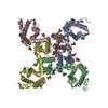
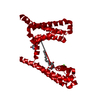
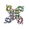
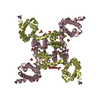
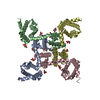
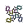
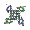
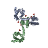
| Title | NaChBac in GDN | ||||||
 Components Components | BH1501 protein | ||||||
 Keywords Keywords | MEMBRANE PROTEIN / NaChBac / Channels / Sodium Ion-Selective | ||||||
| Function / homology |  Function and homology information Function and homology informationvoltage-gated sodium channel complex / voltage-gated sodium channel activity Similarity search - Function | ||||||
| Biological species |  Bacillus halodurans C-125 (bacteria) Bacillus halodurans C-125 (bacteria) | ||||||
| Method | ELECTRON MICROSCOPY / single particle reconstruction / cryo EM / Resolution: 3.7 Å | ||||||
 Authors Authors | Yan, N. / Gao, S. | ||||||
| Funding support |  United States, 1items United States, 1items
| ||||||
 Citation Citation |  Journal: Proc Natl Acad Sci U S A / Year: 2020 Journal: Proc Natl Acad Sci U S A / Year: 2020Title: Employing NaChBac for cryo-EM analysis of toxin action on voltage-gated Na channels in nanodisc. Authors: Shuai Gao / William C Valinsky / Nguyen Cam On / Patrick R Houlihan / Qian Qu / Lei Liu / Xiaojing Pan / David E Clapham / Nieng Yan /   Abstract: NaChBac, the first bacterial voltage-gated Na (Na) channel to be characterized, has been the prokaryotic prototype for studying the structure-function relationship of Na channels. Discovered nearly ...NaChBac, the first bacterial voltage-gated Na (Na) channel to be characterized, has been the prokaryotic prototype for studying the structure-function relationship of Na channels. Discovered nearly two decades ago, the structure of NaChBac has not been determined. Here we present the single particle electron cryomicroscopy (cryo-EM) analysis of NaChBac in both detergent micelles and nanodiscs. Under both conditions, the conformation of NaChBac is nearly identical to that of the potentially inactivated NaAb. Determining the structure of NaChBac in nanodiscs enabled us to examine gating modifier toxins (GMTs) of Na channels in lipid bilayers. To study GMTs in mammalian Na channels, we generated a chimera in which the extracellular fragment of the S3 and S4 segments in the second voltage-sensing domain from Na1.7 replaced the corresponding sequence in NaChBac. Cryo-EM structures of the nanodisc-embedded chimera alone and in complex with HuwenToxin IV (HWTX-IV) were determined to 3.5 and 3.2 Å resolutions, respectively. Compared to the structure of HWTX-IV-bound human Na1.7, which was obtained at an overall resolution of 3.2 Å, the local resolution of the toxin has been improved from ∼6 to ∼4 Å. This resolution enabled visualization of toxin docking. NaChBac can thus serve as a convenient surrogate for structural studies of the interactions between GMTs and Na channels in a membrane environment. | ||||||
| History |
|
- Structure visualization
| Movie |
 Movie viewer Movie viewer |
|---|---|
| Structure viewer | Molecule:  Molmil Molmil Jmol/JSmol Jmol/JSmol |
- Downloads & links
- Download
| PDBx/mmCIF format |  6vx3.cif.gz 6vx3.cif.gz | 177 KB | Display |  PDBx/mmCIF format PDBx/mmCIF format |
|---|---|---|---|---|
| PDB format |  pdb6vx3.ent.gz pdb6vx3.ent.gz | 144.2 KB | Display |  PDB format PDB format |
| PDBx/mmJSON format |  6vx3.json.gz 6vx3.json.gz | Tree view |  PDBx/mmJSON format PDBx/mmJSON format | |
| Others |  Other downloads Other downloads |
-Validation report
| Summary document |  6vx3_validation.pdf.gz 6vx3_validation.pdf.gz | 961.8 KB | Display |  wwPDB validaton report wwPDB validaton report |
|---|---|---|---|---|
| Full document |  6vx3_full_validation.pdf.gz 6vx3_full_validation.pdf.gz | 1012.3 KB | Display | |
| Data in XML |  6vx3_validation.xml.gz 6vx3_validation.xml.gz | 41.3 KB | Display | |
| Data in CIF |  6vx3_validation.cif.gz 6vx3_validation.cif.gz | 50.4 KB | Display | |
| Arichive directory |  https://data.pdbj.org/pub/pdb/validation_reports/vx/6vx3 https://data.pdbj.org/pub/pdb/validation_reports/vx/6vx3 ftp://data.pdbj.org/pub/pdb/validation_reports/vx/6vx3 ftp://data.pdbj.org/pub/pdb/validation_reports/vx/6vx3 | HTTPS FTP |
-Related structure data
| Related structure data |  21428MC  6vwxC  6vxoC  6w6oC M: map data used to model this data C: citing same article ( |
|---|---|
| Similar structure data |
- Links
Links
- Assembly
| Deposited unit | 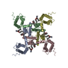
|
|---|---|
| 1 |
|
- Components
| #1: Protein | Mass: 31486.197 Da / Num. of mol.: 4 Source method: isolated from a genetically manipulated source Source: (gene. exp.)  Bacillus halodurans C-125 (bacteria) Bacillus halodurans C-125 (bacteria)Strain: ATCC BAA-125 / DSM 18197 / FERM 7344 / JCM 9153 / C-125 Gene: BH1501 / Production host:  #2: Chemical | ChemComp-POV / ( Has ligand of interest | Y | Has protein modification | N | |
|---|
-Experimental details
-Experiment
| Experiment | Method: ELECTRON MICROSCOPY |
|---|---|
| EM experiment | Aggregation state: PARTICLE / 3D reconstruction method: single particle reconstruction |
- Sample preparation
| Component | Name: NaChBac / Type: COMPLEX / Entity ID: #1 / Source: RECOMBINANT | |||||||||||||||
|---|---|---|---|---|---|---|---|---|---|---|---|---|---|---|---|---|
| Source (natural) | Organism:  Bacillus halodurans C-125 (bacteria) Bacillus halodurans C-125 (bacteria) | |||||||||||||||
| Source (recombinant) | Organism:  | |||||||||||||||
| Buffer solution | pH: 10 | |||||||||||||||
| Buffer component |
| |||||||||||||||
| Specimen | Conc.: 5 mg/ml / Embedding applied: NO / Shadowing applied: NO / Staining applied: NO / Vitrification applied: YES / Details: NaChBac was in lipid nanodisc | |||||||||||||||
| Vitrification | Instrument: FEI VITROBOT MARK IV / Cryogen name: ETHANE / Humidity: 100 % / Chamber temperature: 281 K / Details: blot for 5 seconds before plunging |
- Electron microscopy imaging
| Experimental equipment |  Model: Titan Krios / Image courtesy: FEI Company |
|---|---|
| Microscopy | Model: FEI TITAN KRIOS |
| Electron gun | Electron source:  FIELD EMISSION GUN / Accelerating voltage: 300 kV / Illumination mode: FLOOD BEAM FIELD EMISSION GUN / Accelerating voltage: 300 kV / Illumination mode: FLOOD BEAM |
| Electron lens | Mode: BRIGHT FIELD |
| Image recording | Electron dose: 53 e/Å2 / Film or detector model: GATAN K2 SUMMIT (4k x 4k) |
- Processing
| CTF correction | Type: PHASE FLIPPING AND AMPLITUDE CORRECTION |
|---|---|
| 3D reconstruction | Resolution: 3.7 Å / Resolution method: FSC 0.143 CUT-OFF / Num. of particles: 91616 / Symmetry type: POINT |
 Movie
Movie Controller


 PDBj
PDBj
