 Movie
Movie Controller


+ Open data

- Basic information
| Entry | Database: PDB / ID: 5kmh | |||||||||
|---|---|---|---|---|---|---|---|---|---|---|
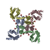
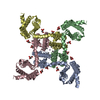
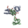
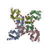
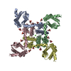
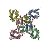
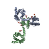
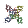
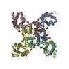
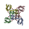
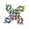
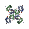
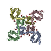
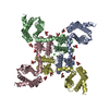
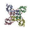
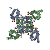
| Title | Structure of CavAb in complex with Br-verapamil | |||||||||
 Components Components | Ion transport protein | |||||||||
 Keywords Keywords | TRANSPORT PROTEIN / Voltage-gated Calcium Channel | |||||||||
| Function / homology |  Function and homology information Function and homology informationvoltage-gated sodium channel complex / voltage-gated sodium channel activity / metal ion binding / identical protein binding Similarity search - Function | |||||||||
| Biological species |  Arcobacter butzleri (bacteria) Arcobacter butzleri (bacteria) | |||||||||
| Method |  X-RAY DIFFRACTION / SYNCHROTRON / MOLECULAR REPLACEMENT / Resolution: 3.2 Å X-RAY DIFFRACTION / SYNCHROTRON / MOLECULAR REPLACEMENT / Resolution: 3.2 Å | |||||||||
 Authors Authors | Tang, L. / Gamal EL-Din, T.M. / Swanson, T.M. / Pryde, D.C. / Scheuer, T. / Zheng, N. / Catterall, W.A. | |||||||||
| Funding support |  United States, 2items United States, 2items
| |||||||||
 Citation Citation | Journal: Nature / Year: 2016 Title: Structural basis for inhibition of a voltage-gated Ca(2+) channel by Ca(2+) antagonist drugs. Authors: Tang, L. / El-Din, T.M. / Swanson, T.M. / Pryde, D.C. / Scheuer, T. / Zheng, N. / Catterall, W.A. | |||||||||
| History |
|
- Structure visualization
| Structure viewer | Molecule: MolmilJmol/JSmol |
|---|
- Downloads & links
-Download
| PDBx/mmCIF format | 5kmh.cif.gz | 397.2 KB | Display | PDBx/mmCIF format |
|---|---|---|---|---|
| PDB format | pdb5kmh.ent.gz | 322.5 KB | Display | PDB format |
| PDBx/mmJSON format | 5kmh.json.gz | Tree view | PDBx/mmJSON format | |
| Others |  Other downloads Other downloads |
-Validation report
| Arichive directory | https://data.pdbj.org/pub/pdb/validation_reports/km/5kmhftp://data.pdbj.org/pub/pdb/validation_reports/km/5kmh | HTTPS FTP |
|---|
-Related structure data
| Related structure data |  5klbC  5klgC  5klsC  5kmdC  5kmfC  4ms2S S: Starting model for refinement C: citing same article ( |
|---|---|
| Similar structure data |
-Links
 PDBj
PDBj
- Assembly
| Deposited unit | 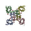
| ||||||||
|---|---|---|---|---|---|---|---|---|---|
| 1 |
| ||||||||
| Unit cell |
|
-Components
| #1: Protein | Mass: 33112.000 Da / Num. of mol.: 4 Source method: isolated from a genetically manipulated source Source: (gene. exp.) Arcobacter butzleri (strain RM4018) (bacteria)Strain: RM4018 / Gene: Abu_1752 / Plasmid: pFASTBAC DUAL / Production host:  Trichoplusia ni (cabbage looper) / References: UniProt: A8EVM5 Trichoplusia ni (cabbage looper) / References: UniProt: A8EVM5#2: Chemical |   Mass: 40.078 Da / Num. of mol.: 3 / Source method: obtained synthetically / Formula: Ca Mass: 40.078 Da / Num. of mol.: 3 / Source method: obtained synthetically / Formula: Ca#3: Chemical | ChemComp-MC3 /   Mass: 677.933 Da / Num. of mol.: 6 / Source method: obtained synthetically / Formula: C36H72NO8P / Comment: phospholipid*YM Mass: 677.933 Da / Num. of mol.: 6 / Source method: obtained synthetically / Formula: C36H72NO8P / Comment: phospholipid*YM#4: Chemical | ChemComp-PX6 /   Mass: 647.883 Da / Num. of mol.: 4 / Source method: obtained synthetically / Formula: C35H68O8P Mass: 647.883 Da / Num. of mol.: 4 / Source method: obtained synthetically / Formula: C35H68O8P#5: Chemical | ChemComp-6U8 / ( |   Mass: 473.446 Da / Num. of mol.: 1 / Source method: obtained synthetically / Formula: C25H33BrN2O2 Mass: 473.446 Da / Num. of mol.: 1 / Source method: obtained synthetically / Formula: C25H33BrN2O2 |
|---|
-Experimental details
-Experiment
| Experiment | Method: X-RAY DIFFRACTION / Number of used crystals: 1 |
|---|
- Sample preparation
| Crystal | Density Matthews: 5.71 Å3/Da / Density % sol: 78.46 % |
|---|---|
| Crystal grow | Temperature: 298 K / Method: vapor diffusion, hanging drop / pH: 5 Details: CHAPSO:DMPC BICELLES,0.1M Na-citrate,pH5.0,2M Ammonium Sulfate,100uM Br-verapamil PH range: 4.7-5.3 |
-Data collection
| Diffraction | Mean temperature: 100 K | |||||||||||||||
|---|---|---|---|---|---|---|---|---|---|---|---|---|---|---|---|---|
| Diffraction source | Source: SYNCHROTRON / Site: ALS / Beamline: 8.2.1 / Wavelength: 0.9198 Å | |||||||||||||||
| Detector | Type: ADSC QUANTUM 315r / Detector: CCD / Date: Sep 25, 2015 | |||||||||||||||
| Radiation | Protocol: SINGLE WAVELENGTH / Monochromatic (M) / Laue (L): M / Scattering type: x-ray | |||||||||||||||
| Radiation wavelength | Wavelength: 0.9198 Å / Relative weight: 1 | |||||||||||||||
| Reflection twin |
| |||||||||||||||
| Reflection | Resolution: 3.2→30 Å / Num. obs: 39188 / % possible obs: 97.7 % / Observed criterion σ(F): 1 / Observed criterion σ(I): 1 / Redundancy: 4.9 % / CC1/2: 0.984 / Rmerge(I) obs: 0.188 / Net I/σ(I): 6 | |||||||||||||||
| Reflection shell | Resolution: 3.2→3.37 Å / Redundancy: 5 % / Rmerge(I) obs: 0.861 / Mean I/σ(I) obs: 1.7 / % possible all: 98.8 |
- Processing
| Software |
| ||||||||||||||||||||||||||||||||||||||||||||||||||||||||||||||||||||||||||||||||||||||||||||||||||||||||||||||||||||||||||||||||||||||||||||||||||||||||||||||||||||||||||||||||||||||
|---|---|---|---|---|---|---|---|---|---|---|---|---|---|---|---|---|---|---|---|---|---|---|---|---|---|---|---|---|---|---|---|---|---|---|---|---|---|---|---|---|---|---|---|---|---|---|---|---|---|---|---|---|---|---|---|---|---|---|---|---|---|---|---|---|---|---|---|---|---|---|---|---|---|---|---|---|---|---|---|---|---|---|---|---|---|---|---|---|---|---|---|---|---|---|---|---|---|---|---|---|---|---|---|---|---|---|---|---|---|---|---|---|---|---|---|---|---|---|---|---|---|---|---|---|---|---|---|---|---|---|---|---|---|---|---|---|---|---|---|---|---|---|---|---|---|---|---|---|---|---|---|---|---|---|---|---|---|---|---|---|---|---|---|---|---|---|---|---|---|---|---|---|---|---|---|---|---|---|---|---|---|---|---|
| Refinement | Method to determine structure: MOLECULAR REPLACEMENT Starting model: 4MS2 Resolution: 3.2→29.83 Å / Cor.coef. Fo:Fc: 0.902 / Cor.coef. Fo:Fc free: 0.832 / SU B: 35.028 / SU ML: 0.287 / Cross valid method: THROUGHOUT / ESU R Free: 0.098 / Details: HYDROGENS HAVE BEEN ADDED IN THE RIDING POSITIONS
| ||||||||||||||||||||||||||||||||||||||||||||||||||||||||||||||||||||||||||||||||||||||||||||||||||||||||||||||||||||||||||||||||||||||||||||||||||||||||||||||||||||||||||||||||||||||
| Solvent computation | Ion probe radii: 0.8 Å / Shrinkage radii: 0.8 Å / VDW probe radii: 1.2 Å | ||||||||||||||||||||||||||||||||||||||||||||||||||||||||||||||||||||||||||||||||||||||||||||||||||||||||||||||||||||||||||||||||||||||||||||||||||||||||||||||||||||||||||||||||||||||
| Displacement parameters | Biso mean: 111.632 Å2
| ||||||||||||||||||||||||||||||||||||||||||||||||||||||||||||||||||||||||||||||||||||||||||||||||||||||||||||||||||||||||||||||||||||||||||||||||||||||||||||||||||||||||||||||||||||||
| Refinement step | Cycle: 1 / Resolution: 3.2→29.83 Å
| ||||||||||||||||||||||||||||||||||||||||||||||||||||||||||||||||||||||||||||||||||||||||||||||||||||||||||||||||||||||||||||||||||||||||||||||||||||||||||||||||||||||||||||||||||||||
| Refine LS restraints |
|