Movie
Movie Controller


+ Open data

- Basic information
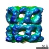
| Entry | Database: EMDB / ID: EMD-20924 | |||||||||
|---|---|---|---|---|---|---|---|---|---|---|
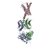
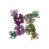
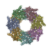
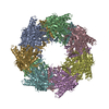
| Title | Single particle cryo-EM structure of KvAP | |||||||||
 Map data Map data | KvAP-6E1 Fab complex | |||||||||
 Sample Sample |
| |||||||||
 Keywords Keywords | voltage-gated potassium channel / non-domain-swapped / TRANSPORT PROTEIN | |||||||||
| Function / homology | Voltage-gated potassium channel / action potential / voltage-gated potassium channel activity / voltage-gated potassium channel complex / Ion transport domain / Ion transport protein / identical protein binding / Voltage-gated potassium channel Function and homology information Function and homology information | |||||||||
| Biological species |   Aeropyrum pernix (archaea) / unidentified (others) Aeropyrum pernix (archaea) / unidentified (others) | |||||||||
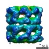
| Method | single particle reconstruction / cryo EM / Resolution: 5.9 Å | |||||||||
 Authors Authors | Tao X / MacKinnon R | |||||||||
| Funding support |  United States, 1 items United States, 1 items
| |||||||||
 Citation Citation | Journal: Elife / Year: 2019 Title: Cryo-EM structure of the KvAP channel reveals a non-domain-swapped voltage sensor topology. Authors: Xiao Tao / Roderick MacKinnon / Abstract: Conductance in voltage-gated ion channels is regulated by membrane voltage through structural domains known as voltage sensors. A single structural class of voltage sensor domain exists, but two ...Conductance in voltage-gated ion channels is regulated by membrane voltage through structural domains known as voltage sensors. A single structural class of voltage sensor domain exists, but two different modes of voltage sensor attachment to the pore occur in nature: domain-swapped and non-domain-swapped. Since the more thoroughly studied Kv1-7, Nav and Cav channels have domain-swapped voltage sensors, much less is known about non-domain-swapped voltage-gated ion channels. In this paper, using cryo-EM, we show that KvAP from has non-domain-swapped voltage sensors as well as other unusual features. The new structure, together with previous functional data, suggests that KvAP and the Shaker channel, to which KvAP is most often compared, probably undergo rather different voltage-dependent conformational changes when they open. | |||||||||
| History |
|
- Structure visualization
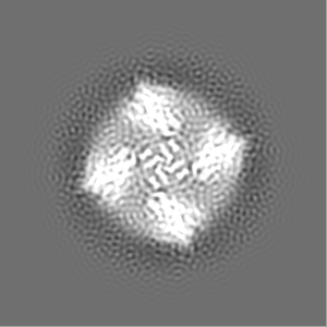
| Movie |
Movie viewer |
|---|---|
| Structure viewer | EM map: SurfViewMolmilJmol/JSmol |
| Supplemental images |

- Downloads & links
-EMDB archive
| Map data | emd_20924.map.gz | 85.4 MB | EMDB map data format | |
|---|---|---|---|---|
| Header (meta data) | emd-20924-v30.xmlemd-20924.xml | 15 KB 15 KB | Display Display | EMDB header |
| Images | 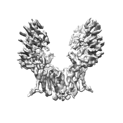 emd_20924.png emd_20924.png | 44.4 KB | ||
| Filedesc metadata | emd-20924.cif.gz | 5.9 KB | ||
| Archive directory |  http://ftp.pdbj.org/pub/emdb/structures/EMD-20924ftp://ftp.pdbj.org/pub/emdb/structures/EMD-20924 http://ftp.pdbj.org/pub/emdb/structures/EMD-20924ftp://ftp.pdbj.org/pub/emdb/structures/EMD-20924 | HTTPS FTP |
-Related structure data
| Related structure data | 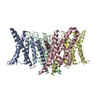 6uwmMC M: atomic model generated by this map C: citing same article ( |
|---|---|
| Similar structure data |
-Links
| EMDB pages | EMDB (EBI/PDBe) / EMDataResource |
|---|---|
| Related items in Molecule of the Month |

-Map
| File | Download / File: emd_20924.map.gz / Format: CCP4 / Size: 103 MB / Type: IMAGE STORED AS FLOATING POINT NUMBER (4 BYTES) | ||||||||||||||||||||||||||||||||||||||||||||||||||||||||||||||||||||
|---|---|---|---|---|---|---|---|---|---|---|---|---|---|---|---|---|---|---|---|---|---|---|---|---|---|---|---|---|---|---|---|---|---|---|---|---|---|---|---|---|---|---|---|---|---|---|---|---|---|---|---|---|---|---|---|---|---|---|---|---|---|---|---|---|---|---|---|---|---|
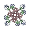
| Annotation | KvAP-6E1 Fab complex | ||||||||||||||||||||||||||||||||||||||||||||||||||||||||||||||||||||
| Projections & slices | Image control
Images are generated by Spider. | ||||||||||||||||||||||||||||||||||||||||||||||||||||||||||||||||||||
| Voxel size | X=Y=Z: 1.028 Å | ||||||||||||||||||||||||||||||||||||||||||||||||||||||||||||||||||||
| Density |
| ||||||||||||||||||||||||||||||||||||||||||||||||||||||||||||||||||||
| Symmetry | Space group: 1 | ||||||||||||||||||||||||||||||||||||||||||||||||||||||||||||||||||||
| Details | EMDB XML:
CCP4 map header:
| ||||||||||||||||||||||||||||||||||||||||||||||||||||||||||||||||||||
 Z (Sec.)
 Y (Row.)
 X (Col.)


-Supplemental data
- Sample components
-Entire : KvAP-6E1 Fab complex
| Entire | Name: KvAP-6E1 Fab complex |
|---|---|
| Components |
|
-Supramolecule #1: KvAP-6E1 Fab complex
| Supramolecule | Name: KvAP-6E1 Fab complex / type: complex / ID: 1 / Parent: 0 / Macromolecule list: all |
|---|---|
| Molecular weight | Theoretical: 200 KDa |
-Supramolecule #2: KvAP
| Supramolecule | Name: KvAP / type: complex / ID: 2 / Parent: 1 / Macromolecule list: all / Details: KvAP tetramer |
|---|---|
| Source (natural) | Organism: Aeropyrum pernix (archaea) |
| Molecular weight | Theoretical: 130 KDa |
-Supramolecule #3: 6E1 Fab
| Supramolecule | Name: 6E1 Fab / type: complex / ID: 3 / Parent: 1 |
|---|---|
| Source (natural) | Organism: unidentified (others) |
-Macromolecule #1: Voltage-gated potassium channel
| Macromolecule | Name: Voltage-gated potassium channel / type: protein_or_peptide / ID: 1 / Number of copies: 4 / Enantiomer: LEVO |
|---|---|
| Source (natural) | Organism: Aeropyrum pernix (archaea) |
| Molecular weight | Theoretical: 31.649207 KDa |
| Recombinant expression | Organism:  |
| Sequence | String: MAGGRVRNIG DVMEHPLVEL GVSYAALLSV IVVVVEYTMQ LSGEYLVRLY LVDLILVIIL WADYAYRAYK SGDPAGYVKK TLYEIPALV PAGLLALIEG HLAGLGLFRL VRLLRFLRIL LIISRGSKFL SAIADAADKI RFYHLFGAVM LTVLYGAFAI Y IVEYPDPN ...String: MAGGRVRNIG DVMEHPLVEL GVSYAALLSV IVVVVEYTMQ LSGEYLVRLY LVDLILVIIL WADYAYRAYK SGDPAGYVKK TLYEIPALV PAGLLALIEG HLAGLGLFRL VRLLRFLRIL LIISRGSKFL SAIADAADKI RFYHLFGAVM LTVLYGAFAI Y IVEYPDPN SSIKSVFDAL WWAVVTATTV GYGDVVPATP IGKVIGIAVM LTGISALTLL IGTVSNMFQK ILVGEPEPSS SP AKLAEMV SSMSEEEFEE FVRTLKNLRR LENSMKLVPR GSRSHHHHHH UniProtKB: Voltage-gated potassium channel |
-Experimental details
-Structure determination
| Method | cryo EM |
|---|---|
 Processing Processing | single particle reconstruction |
| Aggregation state | particle |
-Sample preparation
| Concentration | 6 mg/mL | ||||||||||||
|---|---|---|---|---|---|---|---|---|---|---|---|---|---|
| Buffer | pH: 8 Component:
| ||||||||||||
| Grid | Model: Quantifoil R1.2/1.3 / Material: GOLD / Mesh: 400 / Support film - Material: CARBON / Support film - topology: HOLEY / Pretreatment - Type: GLOW DISCHARGE / Pretreatment - Time: 12 sec. / Pretreatment - Atmosphere: AIR | ||||||||||||
| Vitrification | Cryogen name: ETHANE / Chamber humidity: 100 % / Chamber temperature: 295 K / Instrument: FEI VITROBOT MARK IV / Details: Blot for 4 seconds before plunging.. | ||||||||||||
| Details | KvAP in complex with 6E1 Fab |
- Electron microscopy
| Microscope | FEI TITAN KRIOS |
|---|---|
| Image recording | Film or detector model: GATAN K2 SUMMIT (4k x 4k) / Detector mode: SUPER-RESOLUTION / Digitization - Frames/image: 1-50 / Number grids imaged: 2 / Number real images: 8000 / Average exposure time: 10.0 sec. / Average electron dose: 75.0 e/Å2 |
| Electron beam | Acceleration voltage: 300 kV / Electron source:  FIELD EMISSION GUN FIELD EMISSION GUN |
| Electron optics | C2 aperture diameter: 70.0 µm / Illumination mode: FLOOD BEAM / Imaging mode: BRIGHT FIELD / Cs: 2.7 mm / Nominal defocus max: 2.2 µm / Nominal defocus min: 0.8 µm / Nominal magnification: 29000 |
| Sample stage | Specimen holder model: FEI TITAN KRIOS AUTOGRID HOLDER / Cooling holder cryogen: NITROGEN |
| Experimental equipment |  Model: Titan Krios / Image courtesy: FEI Company |