Movie
Movie Controller

[English] 日本語
 Yorodumi
- EMDB-6725: Folding intermediate of RuBisCO in complex with the GroEL chapero... -
+ Open data

- Basic information
| Entry | Database: EMDB / ID: EMD-6725 | |||||||||
|---|---|---|---|---|---|---|---|---|---|---|
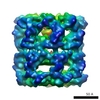
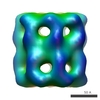
| Title | Folding intermediate of RuBisCO in complex with the GroEL chaperonin. Class1 | |||||||||
 Map data Map data | GroEL:RuBisCO binary complex: Class1 | |||||||||
 Sample Sample |
| |||||||||
| Biological species |  Rhodospirillum rubrum (bacteria) Rhodospirillum rubrum (bacteria) | |||||||||
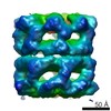
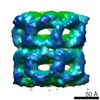
| Method | single particle reconstruction / cryo EM / Resolution: 12.2 Å | |||||||||
 Authors Authors | Natesh R / Clare DK / Farr GW / Horwich AL / Saibil HR | |||||||||
 Citation Citation | Journal: Int J Biol Macromol / Year: 2018 Title: A two-domain folding intermediate of RuBisCO in complex with the GroEL chaperonin. Authors: Ramanathan Natesh / Daniel K Clare / George W Farr / Arthur L Horwich / Helen R Saibil /   Abstract: The chaperonins (GroEL and GroES in Escherichia coli) are ubiquitous molecular chaperones that assist a subset of essential substrate proteins to undergo productive folding to the native state. Using ...The chaperonins (GroEL and GroES in Escherichia coli) are ubiquitous molecular chaperones that assist a subset of essential substrate proteins to undergo productive folding to the native state. Using single particle cryo EM and image processing we have examined complexes of E. coli GroEL with the stringently GroE-dependent substrate enzyme RuBisCO from Rhodospirillum rubrum. Here we present snapshots of non-native RuBisCO - GroEL complexes. We observe two distinct substrate densities in the binary complex reminiscent of the two-domain structure of the RuBisCO subunit, so that this may represent a captured form of an early folding intermediate. The occupancy of the complex is consistent with the negative cooperativity of GroEL with respect to substrate binding, in accordance with earlier mass spectroscopy studies. | |||||||||
| History |
|
- Structure visualization
| Movie |
 Movie viewer Movie viewer |
|---|---|
| Structure viewer | EM map: SurfViewMolmilJmol/JSmol |
| Supplemental images |

- Downloads & links
-EMDB archive
| Map data | emd_6725.map.gz | 1.9 MB | EMDB map data format | |
|---|---|---|---|---|
| Header (meta data) | emd-6725-v30.xmlemd-6725.xml | 17.8 KB 17.8 KB | Display Display | EMDB header |
| Images |  emd_6725.png emd_6725.png | 100.5 KB | ||
| Archive directory |  http://ftp.pdbj.org/pub/emdb/structures/EMD-6725ftp://ftp.pdbj.org/pub/emdb/structures/EMD-6725 http://ftp.pdbj.org/pub/emdb/structures/EMD-6725ftp://ftp.pdbj.org/pub/emdb/structures/EMD-6725 | HTTPS FTP |
-Related structure data


-Links
| EMDB pages | EMDB (EBI/PDBe) / EMDataResource |
|---|
-Map
| File | Download / File: emd_6725.map.gz / Format: CCP4 / Size: 28.7 MB / Type: IMAGE STORED AS FLOATING POINT NUMBER (4 BYTES) | ||||||||||||||||||||||||||||||||||||||||||||||||||||||||||||
|---|---|---|---|---|---|---|---|---|---|---|---|---|---|---|---|---|---|---|---|---|---|---|---|---|---|---|---|---|---|---|---|---|---|---|---|---|---|---|---|---|---|---|---|---|---|---|---|---|---|---|---|---|---|---|---|---|---|---|---|---|---|
| Annotation | GroEL:RuBisCO binary complex: Class1 | ||||||||||||||||||||||||||||||||||||||||||||||||||||||||||||
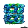
| Projections & slices | Image control
Images are generated by Spider. | ||||||||||||||||||||||||||||||||||||||||||||||||||||||||||||
| Voxel size | X=Y=Z: 2.8 Å | ||||||||||||||||||||||||||||||||||||||||||||||||||||||||||||
| Density |
| ||||||||||||||||||||||||||||||||||||||||||||||||||||||||||||
| Symmetry | Space group: 1 | ||||||||||||||||||||||||||||||||||||||||||||||||||||||||||||
| Details | EMDB XML:
CCP4 map header:
| ||||||||||||||||||||||||||||||||||||||||||||||||||||||||||||
 Z (Sec.)
 Y (Row.)
 X (Col.)


-Supplemental data
- Sample components
-Entire : Non-native RuBisCO in complex with chaperonin GroEL
| Entire | Name: Non-native RuBisCO in complex with chaperonin GroEL |
|---|---|
| Components |
|
-Supramolecule #1: Non-native RuBisCO in complex with chaperonin GroEL
| Supramolecule | Name: Non-native RuBisCO in complex with chaperonin GroEL / type: complex / ID: 1 / Parent: 0 / Macromolecule list: all / Details: GroEL.D473C.His6 with RuBisCO folding intermediate |
|---|---|
| Source (natural) | Organism: |
| Recombinant expression | Organism: |
| Molecular weight | Theoretical: 863 KDa |
-Macromolecule #1: GroEL
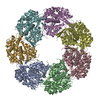
| Macromolecule | Name: GroEL / type: protein_or_peptide / ID: 1 / Details: GroEL tetradecamer. / Enantiomer: LEVO / EC number: ec: 3.6.4.9 |
|---|---|
| Source (natural) | Organism: |
| Recombinant expression | Organism: |
| Sequence | String: AAKDVKFGND AGVKMLRGVN VLADAVKVTL GPKGRNVVLD KSFGAPTITK DGVSVAREIE LEDKFENMGA QMVKEVASKA NDAAGDGTTT ATVLAQAIIT EGLKAVAAGM NPMDLKRGID KAVTVAVEEL KALSVPCSDS KAIAQVGTIS ANSDETVGKL IAEAMDKVGK ...String: AAKDVKFGND AGVKMLRGVN VLADAVKVTL GPKGRNVVLD KSFGAPTITK DGVSVAREIE LEDKFENMGA QMVKEVASKA NDAAGDGTTT ATVLAQAIIT EGLKAVAAGM NPMDLKRGID KAVTVAVEEL KALSVPCSDS KAIAQVGTIS ANSDETVGKL IAEAMDKVGK EGVITVEDGT GLQDELDVVE GMQFDRGYLS PYFINKPETG AVELESPFIL LADKKISNIR EMLPVLEAVA KAGKPLLIIA EDVEGEALAT AVVNTIRGIV KVAAVKAPGF GDRRKAMLQD IATLTGGTVI SEEIGMELEK ATLEDLGQAK RVVINKDTTT IIDGVGEEAA IQGRVAQIRQ QIEEATSDYD REKLQERVAK LAGGVAVIKV GAATEVEMKE KKARVEDALH ATRAAVEEGV VAGGGVALIR VASKLADLRG QNEDQNVGIK VALRAMEAPL RQIVLNCGEE PSVVANTVKG GCGNYGYNAA TEEYGNMIDM GILDPTKVTR SALQYAASVA GLMITTECMV TDLP |
-Macromolecule #2: RuBisCO
| Macromolecule | Name: RuBisCO / type: protein_or_peptide / ID: 2 / Details: RuBisCO / Enantiomer: LEVO / EC number: ribulose-bisphosphate carboxylase |
|---|---|
| Source (natural) | Organism: Rhodospirillum rubrum (bacteria) |
| Recombinant expression | Organism: |
| Sequence | String: TMITNSPDRW GYSAPHRTSR ESPPMDQSSR YVNLALKEED LIAGGEHVLC AYIMKPKAGY GYVATAAHFA AESSTGTNVE VCTTDDFTRG VDALVYEVDE ARELTKIAYP VALFDRNITD GKAMIASFLT LTMGNNQGMG DVEYAKMHDF YVPEAYRALF DGPSVNISAL ...String: TMITNSPDRW GYSAPHRTSR ESPPMDQSSR YVNLALKEED LIAGGEHVLC AYIMKPKAGY GYVATAAHFA AESSTGTNVE VCTTDDFTRG VDALVYEVDE ARELTKIAYP VALFDRNITD GKAMIASFLT LTMGNNQGMG DVEYAKMHDF YVPEAYRALF DGPSVNISAL WKVLGRPEVD GGLVVGTIIK PKLGLRPKPF AEACHAFWLG GDFIKNDEPQ GNQPFAPLRD TIALVADAMR RAQDETGEAK LFSANITADD PFEIIARGEY VLETFGENAS HVALLVDGYV AGAAAITTAR RRFPDNFLHY HRAGHGAVTS PQSKRGYTAF VHCKMARLQG ASGIHTGTMG FGKMEGESSD RAIAYMLTQD EAQGPFYRQS WGGMKACTPI ISGGMNALRM PGFFENLGNA NVILTAGGGA FGHIDGPVAG ARSLRQAWQA WRDGVPVLDY AREHKELARA FESFPGDADQ IYPGWRKALG VEDTRSALPA |
-Experimental details
-Structure determination
| Method | cryo EM |
|---|---|
 Processing Processing | single particle reconstruction |
| Aggregation state | particle |
-Sample preparation
| Concentration | 0.0863 mg/mL | |||||||||||||||
|---|---|---|---|---|---|---|---|---|---|---|---|---|---|---|---|---|
| Buffer | pH: 7.5 Component:
Details: Folding Buffer (FB) consists of 50 mM HEPES pH7.5, 10 mM KOAc, 10 mM Mg(OAc)2, 10 mM DTT. RuBisCO denatured in 20mM HCl, 10 M Urea, 20 mM DTT | |||||||||||||||
| Grid | Model: Protochips Inc., USA / Material: COPPER / Mesh: 300 / Support film - Material: CARBON / Support film - topology: HOLEY / Pretreatment - Type: GLOW DISCHARGE / Pretreatment - Atmosphere: AIR Details: The C-flat holey grids were coated with thin home made carbon film. | |||||||||||||||
| Vitrification | Cryogen name: ETHANE / Chamber temperature: 298 K / Instrument: HOMEMADE PLUNGER / Details: Blot approx. 1 sec before plunging. Visual.. | |||||||||||||||
| Details | RuBisCO denatured in acid urea was complexed with GroEL.D473C.His6 in Folding Buffer |
- Electron microscopy
| Microscope | FEI TECNAI F20 |
|---|---|
| Temperature | Min: 100.0 K / Max: 100.0 K |
| Image recording | Film or detector model: KODAK SO-163 FILM / Digitization - Sampling interval: 7.0 µm / Number grids imaged: 11 / Number real images: 468 / Average exposure time: 1.0 sec. / Average electron dose: 10.0 e/Å2 Details: Images were collected on Kodak SO-163 Film. Dose was 10-15 e-/A2/sec. |
| Electron beam | Acceleration voltage: 200 kV / Electron source:  FIELD EMISSION GUN FIELD EMISSION GUN |
| Electron optics | C2 aperture diameter: 40.0 µm / Calibrated magnification: 50000 / Illumination mode: FLOOD BEAM / Imaging mode: BRIGHT FIELD / Cs: 2.0 mm / Nominal defocus max: 3.5 µm / Nominal defocus min: 1.0 µm / Nominal magnification: 50000 |
| Sample stage | Specimen holder model: GATAN CT3500 SINGLE TILT LIQUID NITROGEN CRYO TRANSFER HOLDER Cooling holder cryogen: NITROGEN |
| Experimental equipment |  Model: Tecnai F20 / Image courtesy: FEI Company |
+Image processing
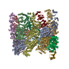
-Atomic model buiding 1
| Initial model | PDB ID: Chain - Residue range: 2-525 |
|---|---|
| Details | Rigid body fit |
| Refinement | Protocol: RIGID BODY FIT |