Movie
Movie Controller

[English] 日本語
 Yorodumi
- EMDB-1544: Structure of a chaperonin complex with non-native substrate prote... -
+ Open data

- Basic information
| Entry | Database: EMDB / ID: EMD-1544 | |||||||||
|---|---|---|---|---|---|---|---|---|---|---|
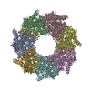
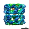
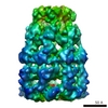
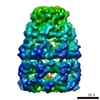
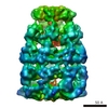
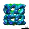
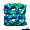
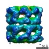
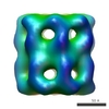
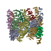
| Title | Structure of a chaperonin complex with non-native substrate protein gp23 | |||||||||
 Map data Map data | Volume contains GroEL complexed with denatured gp23 without imposed symmetry | |||||||||
 Sample Sample |
| |||||||||
 Keywords Keywords | chaperonin / bacteriophage T4 / capsid protein gp23 / GroEL | |||||||||
| Function / homology | Capsid protein, T4-like bacteriophage-like / protein binding / Chaperonin Cpn60/GroEL Function and homology information Function and homology information | |||||||||
| Biological species |   Enterobacteria phage T4 (virus) Enterobacteria phage T4 (virus) | |||||||||
| Method | single particle reconstruction / cryo EM / negative staining / Resolution: 10.7 Å | |||||||||
 Authors Authors | Clare DK / Bakkes PJ / van Heerikhuizen H / van der Vies SM / Saibil HR | |||||||||
 Citation Citation | Journal: Nature / Year: 2009 Title: Chaperonin complex with a newly folded protein encapsulated in the folding chamber. Authors: D K Clare / P J Bakkes / H van Heerikhuizen / S M van der Vies / H R Saibil /  Abstract: A subset of essential cellular proteins requires the assistance of chaperonins (in Escherichia coli, GroEL and GroES), double-ring complexes in which the two rings act alternately to bind, ...A subset of essential cellular proteins requires the assistance of chaperonins (in Escherichia coli, GroEL and GroES), double-ring complexes in which the two rings act alternately to bind, encapsulate and fold a wide range of nascent or stress-denatured proteins. This process starts by the trapping of a substrate protein on hydrophobic surfaces in the central cavity of a GroEL ring. Then, binding of ATP and co-chaperonin GroES to that ring ejects the non-native protein from its binding sites, through forced unfolding or other major conformational changes, and encloses it in a hydrophilic chamber for folding. ATP hydrolysis and subsequent ATP binding to the opposite ring trigger dissociation of the chamber and release of the substrate protein. The bacteriophage T4 requires its own version of GroES, gp31, which forms a taller folding chamber, to fold the major viral capsid protein gp23 (refs 16-20). Polypeptides are known to fold inside the chaperonin complex, but the conformation of an encapsulated protein has not previously been visualized. Here we present structures of gp23-chaperonin complexes, showing both the initial captured state and the final, close-to-native state with gp23 encapsulated in the folding chamber. Although the chamber is expanded, it is still barely large enough to contain the elongated gp23 monomer, explaining why the GroEL-GroES complex is not able to fold gp23 and showing how the chaperonin structure distorts to enclose a large, physiological substrate protein. | |||||||||
| History |
|
- Structure visualization
| Movie |
Movie viewer |
|---|---|
| Structure viewer | EM map: SurfViewMolmilJmol/JSmol |
| Supplemental images |

- Downloads & links
-EMDB archive
| Map data | emd_1544.map.gz | 216.1 KB | EMDB map data format | |
|---|---|---|---|---|
| Header (meta data) | emd-1544-v30.xmlemd-1544.xml | 11.8 KB 11.8 KB | Display Display | EMDB header |
| Images |  1544.gif 1544.gif | 51.5 KB | ||
| Archive directory |  http://ftp.pdbj.org/pub/emdb/structures/EMD-1544ftp://ftp.pdbj.org/pub/emdb/structures/EMD-1544 http://ftp.pdbj.org/pub/emdb/structures/EMD-1544ftp://ftp.pdbj.org/pub/emdb/structures/EMD-1544 | HTTPS FTP |
-Related structure data


-Links
| EMDB pages | EMDB (EBI/PDBe) / EMDataResource |
|---|
-Map
| File | Download / File: emd_1544.map.gz / Format: CCP4 / Size: 15.3 MB / Type: IMAGE STORED AS FLOATING POINT NUMBER (4 BYTES) | ||||||||||||||||||||||||||||||||||||||||||||||||||||||||||||||||||||
|---|---|---|---|---|---|---|---|---|---|---|---|---|---|---|---|---|---|---|---|---|---|---|---|---|---|---|---|---|---|---|---|---|---|---|---|---|---|---|---|---|---|---|---|---|---|---|---|---|---|---|---|---|---|---|---|---|---|---|---|---|---|---|---|---|---|---|---|---|---|
| Annotation | Volume contains GroEL complexed with denatured gp23 without imposed symmetry | ||||||||||||||||||||||||||||||||||||||||||||||||||||||||||||||||||||
| Projections & slices | Image control
Images are generated by Spider. | ||||||||||||||||||||||||||||||||||||||||||||||||||||||||||||||||||||
| Voxel size | X=Y=Z: 2.8 Å | ||||||||||||||||||||||||||||||||||||||||||||||||||||||||||||||||||||
| Density |
| ||||||||||||||||||||||||||||||||||||||||||||||||||||||||||||||||||||
| Symmetry | Space group: 1 | ||||||||||||||||||||||||||||||||||||||||||||||||||||||||||||||||||||
| Details | EMDB XML:
CCP4 map header:
| ||||||||||||||||||||||||||||||||||||||||||||||||||||||||||||||||||||
 Z (Sec.)
 X (Row.)
 Y (Col.)


-Supplemental data
- Sample components
-Entire : GroEL-gp23
| Entire | Name: GroEL-gp23 |
|---|---|
| Components |
|
-Supramolecule #1000: GroEL-gp23
| Supramolecule | Name: GroEL-gp23 / type: sample / ID: 1000 Details: gp23 was denatured in UREA and mixed with GroEL containing buffer Oligomeric state: tetradecamer of GroEL bound to one copy of denatured gp23 Number unique components: 2 |
|---|---|
| Molecular weight | Experimental: 850 KDa / Theoretical: 850 KDa / Method: sequence |
-Macromolecule #1: GroEL
| Macromolecule | Name: GroEL / type: protein_or_peptide / ID: 1 / Name.synonym: GroEL / Number of copies: 1 / Oligomeric state: tetradecamer / Recombinant expression: Yes |
|---|---|
| Source (natural) | Organism: |
| Molecular weight | Experimental: 56 KDa / Theoretical: 56 KDa |
| Recombinant expression | Organism: |
| Sequence | GO: protein binding / InterPro: Chaperonin Cpn60/GroEL |
-Macromolecule #2: gp23
| Macromolecule | Name: gp23 / type: protein_or_peptide / ID: 2 / Name.synonym: Major capsid protein / Number of copies: 1 / Oligomeric state: monomer / Recombinant expression: Yes |
|---|---|
| Source (natural) | Organism: Enterobacteria phage T4 (virus) / synonym: Bacteriophage T4 / Location in cell: cytoplasmic |
| Molecular weight | Experimental: 56 KDa / Theoretical: 56 KDa |
| Recombinant expression | Organism: |
| Sequence | InterPro: Capsid protein, T4-like bacteriophage-like |
-Experimental details
-Structure determination
| Method | negative staining, cryo EM |
|---|---|
 Processing Processing | single particle reconstruction |
| Aggregation state | particle |
-Sample preparation
| Concentration | 1 mg/mL |
|---|---|
| Buffer | pH: 7.5 / Details: 40 mM Tris-HCl pH 7.5, 10 mM KCl, 10 mM MgCl2 |
| Staining | Type: NEGATIVE / Details: Cryo |
| Grid | Details: 400 mesh R2/2 c-flat grids |
| Vitrification | Cryogen name: ETHANE / Chamber temperature: 100 K / Instrument: HOMEMADE PLUNGER / Details: Vitrification instrument: manual plunger / Method: blot for 2-3 seconds before plunging |
- Electron microscopy
| Microscope | FEI TECNAI F20 |
|---|---|
| Temperature | Average: 100 K |
| Alignment procedure | Legacy - Astigmatism: corrected at 150,00 times |
| Date | Jun 1, 2006 |
| Image recording | Category: FILM / Film or detector model: KODAK SO-163 FILM / Digitization - Scanner: ZEISS SCAI / Digitization - Sampling interval: 1.4 µm / Number real images: 150 / Average electron dose: 15 e/Å2 / Od range: 0.5 / Bits/pixel: 8 |
| Electron beam | Acceleration voltage: 200 kV / Electron source:  FIELD EMISSION GUN FIELD EMISSION GUN |
| Electron optics | Calibrated magnification: 50000 / Illumination mode: FLOOD BEAM / Imaging mode: BRIGHT FIELD / Cs: 2 mm / Nominal defocus max: 3.0 µm / Nominal defocus min: 1.0 µm / Nominal magnification: 50000 |
| Sample stage | Specimen holder: side entry / Specimen holder model: GATAN LIQUID NITROGEN |
| Experimental equipment |  Model: Tecnai F20 / Image courtesy: FEI Company |
-Image processing
| Details | No symmetry was applied |
|---|---|
| CTF correction | Details: Each particle |
| Final reconstruction | Applied symmetry - Point group: C1 (asymmetric) / Algorithm: OTHER / Resolution.type: BY AUTHOR / Resolution: 10.7 Å / Resolution method: FSC 0.5 CUT-OFF / Software - Name: SPIDER / Details: No symmetry was applied to the reconstruction / Number images used: 5600 |
| Final angle assignment | Details: SPIDER:theta 78-90 degrees, phi 0-360 degrees. Mirror option was on. |
-Atomic model buiding 1
| Initial model | PDB ID: |
|---|---|
| Software | Name: URO |
| Details | Protocol: Individual domains fitted as rigid bodies. The domains were separately fitted in URO |
| Refinement | Space: RECIPROCAL / Protocol: RIGID BODY FIT / Target criteria: cross-correlation coefficient |