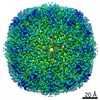
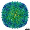
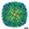
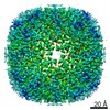
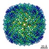
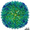
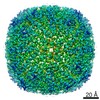
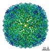
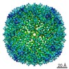
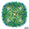
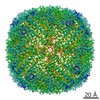
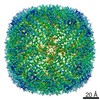
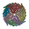
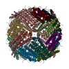
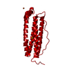
- PDB-6sht: Molecular structure of mouse apoferritin resolved at 2.7 Angstrom... -
+
データを開く
IDまたはキーワード:
読み込み中...
-
基本情報
登録情報
データベース: PDB / ID: 6sht
タイトル
Molecular structure of mouse apoferritin resolved at 2.7 Angstroms with the Glacios cryo-microscope
要素
Ferritin heavy chain
キーワード
METAL BINDING PROTEIN / Apoferritin / iron binding / iron storing / complex
機能・相同性
機能・相同性情報
Iron uptake and transport / Golgi Associated Vesicle Biogenesis / iron ion sequestering activity / autolysosome / ferroxidase / intracellular sequestering of iron ion / ferroxidase activity / negative regulation of fibroblast proliferation / endocytic vesicle lumen / Neutrophil degranulation ...Iron uptake and transport / Golgi Associated Vesicle Biogenesis / iron ion sequestering activity / autolysosome / ferroxidase / intracellular sequestering of iron ion / ferroxidase activity / negative regulation of fibroblast proliferation / endocytic vesicle lumen / Neutrophil degranulation / ferric iron binding / ferrous iron binding / iron ion transport / iron ion binding / immune response / negative regulation of cell population proliferation / mitochondrion / extracellular region / identical protein binding / membrane / cytoplasm / cytosol 類似検索 - 分子機能
ジャーナル: PLoS One / 年: 2020 タイトル: 2.7 Å cryo-EM structure of vitrified M. musculus H-chain apoferritin from a compact 200 keV cryo-microscope. 著者: Farzad Hamdi / Christian Tüting / Dmitry A Semchonok / Koen M Visscher / Fotis L Kyrilis / Annette Meister / Ioannis Skalidis / Lisa Schmidt / Christoph Parthier / Milton T Stubbs / Panagiotis L Kastritis / 要旨: Here we present the structure of mouse H-chain apoferritin at 2.7 Å (FSC = 0.143) solved by single particle cryogenic electron microscopy (cryo-EM) using a 200 kV device, the Thermo Fisher Glacios®. ...Here we present the structure of mouse H-chain apoferritin at 2.7 Å (FSC = 0.143) solved by single particle cryogenic electron microscopy (cryo-EM) using a 200 kV device, the Thermo Fisher Glacios®. This is a compact, two-lens illumination system with a constant power objective lens, without any energy filters or aberration correctors, often thought of as a "screening cryo-microscope". Coulomb potential maps reveal clear densities for main chain carbonyl oxygens, residue side chains (including alternative conformations) and bound solvent molecules. We used a quasi-crystallographic reciprocal space approach to fit model coordinates to the experimental cryo-EM map. We argue that the advantages offered by (a) the high electronic and mechanical stability of the microscope, (b) the high emission stability and low beam energy spread of the high brightness Field Emission Gun (X-FEG), (c) direct electron detection technology and (d) particle-based Contrast Transfer Function (CTF) refinement have contributed to achieving high resolution. Overall, we show that basic electron optical settings for automated cryo-electron microscopy imaging can be used to determine structures approaching atomic resolution.
 ムービー
ムービー コントローラー


 データを開く
 基本情報
基本情報 要素
要素 キーワード
キーワード 機能・相同性情報
 データ登録者
 引用
引用

 構造の表示
構造の表示 ダウンロードとリンク
 その他のダウンロード


 PDBj
PDBj


 集合体
集合体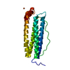


 分子量: 55.845 Da / 分子数: 1 / 由来タイプ: 合成 / 式: Fe / タイプ: SUBJECT OF INVESTIGATION
 分子量: 24.305 Da / 分子数: 1 / 由来タイプ: 合成 / 式: Mg / タイプ: SUBJECT OF INVESTIGATION
 分子量: 18.015 Da / 分子数: 72 / 由来タイプ: 天然 / 式: H2O
 試料調製
試料調製 電子顕微鏡撮影
 FIELD EMISSION GUN / 加速電圧: 200 kV / 照射モード: FLOOD BEAM
 解析
解析