[English] 日本語
Yorodumi
- PDB-6hiz: Cryo-EM structure of the Trypanosoma brucei mitochondrial ribosom... -

+
Open data


ID or keywords:

Loading...

-
Basic information

Entry
Database: PDB / ID: 6hiz
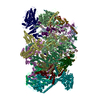
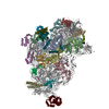
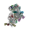
TitleCryo-EM structure of the Trypanosoma brucei mitochondrial ribosome - This entry contains the head of the small mitoribosomal subunit
Components
  • (Unknown protein) x 2
  • RNA (143-MER)
  • mS29
  • mS33
  • mS35
  • mS48
  • mS49
  • mS50
  • mS52
  • mS53
  • mS54
  • mS55
  • mS57
  • mS58
  • mS59
  • mS67
  • mS69
  • mS70
  • mS71
  • mS72
  • uS10m
  • uS11m
  • uS14m
  • uS18m
  • uS19m
  • uS3m
  • uS9m
KeywordsRIBOSOME / mitoribosome / translation / Trypanosoma / small ribosomal subunit / 9S rRNA / ribosomal protein
Function / homology
Function and homology information


mitochondrial mRNA editing complex / mitochondrial RNA processing / thiosulfate sulfurtransferase activity / kinetoplast / mRNA stabilization / nuclear lumen / ciliary plasm / mitochondrial small ribosomal subunit / structural constituent of ribosome / translation ...mitochondrial mRNA editing complex / mitochondrial RNA processing / thiosulfate sulfurtransferase activity / kinetoplast / mRNA stabilization / nuclear lumen / ciliary plasm / mitochondrial small ribosomal subunit / structural constituent of ribosome / translation / mitochondrion / RNA binding / nucleoplasm / nucleus / cytoplasm
Similarity search - Function
Ribosomal protein S23/S29, mitochondrial / Mitochondrial ribosomal death-associated protein 3 / Rhodanese-like domain / LysM domain / Rhodanese domain profile. / Rhodanese-like domain superfamily / LysM domain / Rhodanese-like domain / Ribosomal protein S18 superfamily / Ribosomal protein S9 ...Ribosomal protein S23/S29, mitochondrial / Mitochondrial ribosomal death-associated protein 3 / Rhodanese-like domain / LysM domain / Rhodanese domain profile. / Rhodanese-like domain superfamily / LysM domain / Rhodanese-like domain / Ribosomal protein S18 superfamily / Ribosomal protein S9 / Ribosomal protein S9/S16 / Ribosomal protein S11 superfamily / Tetratricopeptide-like helical domain superfamily / Ribosomal protein S5 domain 2-type fold, subgroup / Ribosomal protein S5 domain 2-type fold
Similarity search - Domain/homology
GUANOSINE-5'-TRIPHOSPHATE / SPERMIDINE / URIDINE 5'-TRIPHOSPHATE / : / RNA / RNA (> 10) / RNA (> 100) / Uncharacterized protein / Uncharacterized protein / Mitochondrial SSU ribosomal protein ...GUANOSINE-5'-TRIPHOSPHATE / SPERMIDINE / URIDINE 5'-TRIPHOSPHATE / : / RNA / RNA (> 10) / RNA (> 100) / Uncharacterized protein / Uncharacterized protein / Mitochondrial SSU ribosomal protein / Mitochondrial SSU ribosomal protein / Uncharacterized protein / Mitochondrial ribosomal protein S18 / Uncharacterized protein / Uncharacterized protein / Uncharacterized protein / Uncharacterized protein / Uncharacterized protein / Ribosomal protein S9 / Uncharacterized protein / Uncharacterized protein / Uncharacterized protein / LysM domain-containing protein / Uncharacterized protein / Uncharacterized protein / Pentacotripeptide-repeat region of PRORP domain-containing protein / Uncharacterized protein / Uncharacterized protein / Small ribosomal subunit protein mS29 / Rhodanese domain-containing protein / Uncharacterized protein
Similarity search - Component
Biological speciesTrypanosoma brucei brucei (eukaryote)
MethodELECTRON MICROSCOPY / single particle reconstruction / cryo EM / Resolution: 3.08 Å
AuthorsRamrath, D.J.F. / Niemann, M. / Leibundgut, M. / Bieri, P. / Prange, C. / Horn, E.K. / Leitner, A. / Boehringer, A. / Schneider, A. / Ban, N.
Funding support Switzerland, 1items
OrganizationGrant numberCountry
Swiss National Science Foundation Switzerland
CitationJournal: Science / Year: 2018
Title: Evolutionary shift toward protein-based architecture in trypanosomal mitochondrial ribosomes.
Authors: David J F Ramrath / Moritz Niemann / Marc Leibundgut / Philipp Bieri / Céline Prange / Elke K Horn / Alexander Leitner / Daniel Boehringer / André Schneider / Nenad Ban /
Abstract: Ribosomal RNA (rRNA) plays key functional and architectural roles in ribosomes. Using electron microscopy, we determined the atomic structure of a highly divergent ribosome found in mitochondria of , ...Ribosomal RNA (rRNA) plays key functional and architectural roles in ribosomes. Using electron microscopy, we determined the atomic structure of a highly divergent ribosome found in mitochondria of , a unicellular parasite that causes sleeping sickness in humans. The trypanosomal mitoribosome features the smallest rRNAs and contains more proteins than all known ribosomes. The structure shows how the proteins have taken over the role of architectural scaffold from the rRNA: They form an autonomous outer shell that surrounds the entire particle and stabilizes and positions the functionally important regions of the rRNA. Our results also reveal the "minimal" set of conserved rRNA and protein components shared by all ribosomes that help us define the most essential functional elements.
History
DepositionAug 31, 2018Deposition site: PDBE / Processing site: PDBE
Revision 1.0Sep 26, 2018Provider: repository / Type: Initial release
Revision 1.1Nov 7, 2018Group: Data collection / Database references / Category: citation / Item: _citation.journal_volume

-
Structure visualization

Movie
  • Deposited structure unit
  • Imaged by Jmol
  • Download
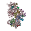
  • Superimposition on EM map
  • EMDB-0233
  • Imaged by UCSF Chimera
  • Download
Movie viewer
Structure viewerMolecule:
MolmilJmol/JSmol

Downloads & links

-
Assembly

Deposited unit
DA: mS48
DL: mS59
DB: mS49
DC: mS50
DE: mS52
DF: mS53
DG: mS54
DH: mS55
DJ: mS57
DK: mS58
DT: mS67
DV: mS69
DW: mS70
DX: mS71
DY: mS72
CC: uS3m
CI: uS9m
CJ: uS10m
CK: uS11m
CN: uS14m
CR: uS18m
CS: uS19m
Cg: mS29
Ci: mS33
Ck: mS35
CA: RNA (143-MER)
UO: Unknown protein
UP: Unknown protein
hetero molecules


Theoretical massNumber of molelcules
Total (without water)1,650,36735
Polymers1,648,99628
Non-polymers1,3717
Water543
1


  • Idetical with deposited unit
  • defined by author
  • Evidence: mass spectrometry
TypeNameSymmetry operationNumber
identity operation1_5551

-
Components

+
Protein , 25 types, 25 molecules DADLDBDCDEDFDGDHDJDKDTDVDWDXDYCCCICJCKCNCRCSCgCiCk

#1: Protein mS48


Mass: 201811.188 Da / Num. of mol.: 1 / Source method: isolated from a natural source / Source: (natural) Trypanosoma brucei brucei (eukaryote) / Variant: Lister 427 / References: UniProt: Q57UJ2
#2: Protein mS59


Mass: 35395.676 Da / Num. of mol.: 1 / Source method: isolated from a natural source / Source: (natural) Trypanosoma brucei brucei (eukaryote) / Variant: Lister 427 / References: UniProt: Q38BS2
#3: Protein mS49


Mass: 137234.125 Da / Num. of mol.: 1 / Source method: isolated from a natural source / Source: (natural) Trypanosoma brucei brucei (eukaryote) / Variant: Lister 427 / References: UniProt: Q586P5
#4: Protein mS50


Mass: 131719.250 Da / Num. of mol.: 1 / Source method: isolated from a natural source / Source: (natural) Trypanosoma brucei brucei (eukaryote) / Variant: Lister 427 / References: UniProt: Q57YB5
#5: Protein mS52


Mass: 85012.258 Da / Num. of mol.: 1 / Source method: isolated from a natural source / Source: (natural) Trypanosoma brucei brucei (eukaryote) / Variant: Lister 427 / References: UniProt: Q386Q7
#6: Protein mS53


Mass: 75338.914 Da / Num. of mol.: 1 / Source method: isolated from a natural source / Source: (natural) Trypanosoma brucei brucei (eukaryote) / Variant: Lister 427 / References: UniProt: Q38ET1
#7: Protein mS54


Mass: 72046.031 Da / Num. of mol.: 1 / Source method: isolated from a natural source / Source: (natural) Trypanosoma brucei brucei (eukaryote) / Variant: Lister 427 / References: UniProt: Q57ZP8
#8: Protein mS55


Mass: 66676.680 Da / Num. of mol.: 1 / Source method: isolated from a natural source / Source: (natural) Trypanosoma brucei brucei (eukaryote) / Variant: Lister 427 / References: UniProt: Q580V1
#9: Protein mS57


Mass: 45956.293 Da / Num. of mol.: 1 / Source method: isolated from a natural source / Source: (natural) Trypanosoma brucei brucei (eukaryote) / Variant: Lister 427 / References: UniProt: Q584U8
#10: Protein mS58


Mass: 35623.910 Da / Num. of mol.: 1 / Source method: isolated from a natural source / Source: (natural) Trypanosoma brucei brucei (eukaryote) / Variant: Lister 427 / References: UniProt: Q38BP1
#11: Protein mS67


Mass: 30096.051 Da / Num. of mol.: 1 / Source method: isolated from a natural source / Source: (natural) Trypanosoma brucei brucei (eukaryote) / Variant: Lister 427 / References: UniProt: Q586G5
#12: Protein mS69


Mass: 21589.732 Da / Num. of mol.: 1 / Source method: isolated from a natural source / Source: (natural) Trypanosoma brucei brucei (eukaryote) / Variant: Lister 427 / References: UniProt: Q57UZ6
#13: Protein mS70


Mass: 21247.398 Da / Num. of mol.: 1 / Source method: isolated from a natural source / Source: (natural) Trypanosoma brucei brucei (eukaryote) / Variant: Lister 427 / References: UniProt: Q383N9
#14: Protein mS71


Mass: 20093.227 Da / Num. of mol.: 1 / Source method: isolated from a natural source / Source: (natural) Trypanosoma brucei brucei (eukaryote) / Variant: Lister 427 / References: UniProt: Q383G5
#15: Protein mS72


Mass: 19317.293 Da / Num. of mol.: 1 / Source method: isolated from a natural source / Source: (natural) Trypanosoma brucei brucei (eukaryote) / Variant: Lister 427 / References: UniProt: Q57YD4
#16: Protein uS3m


Mass: 9070.940 Da / Num. of mol.: 1 / Source method: isolated from a natural source / Details: Mitochondrial gene uS3m (MURF-5) / Source: (natural) Trypanosoma brucei brucei (eukaryote) / Variant: Lister 427
#17: Protein uS9m


Mass: 50128.926 Da / Num. of mol.: 1 / Source method: isolated from a natural source / Source: (natural) Trypanosoma brucei brucei (eukaryote) / Variant: Lister 427 / References: UniProt: Q57W62
#18: Protein uS10m


Mass: 94351.039 Da / Num. of mol.: 1 / Source method: isolated from a natural source / Source: (natural) Trypanosoma brucei brucei (eukaryote) / Variant: Lister 427 / References: UniProt: Q57Z45
#19: Protein uS11m


Mass: 37925.910 Da / Num. of mol.: 1 / Source method: isolated from a natural source / Source: (natural) Trypanosoma brucei brucei (eukaryote) / Variant: Lister 427 / References: UniProt: Q389T7
#20: Protein uS14m


Mass: 19900.014 Da / Num. of mol.: 1 / Source method: isolated from a natural source / Source: (natural) Trypanosoma brucei brucei (eukaryote) / Variant: Lister 427 / References: UniProt: Q580I0
#21: Protein uS18m


Mass: 36988.691 Da / Num. of mol.: 1 / Source method: isolated from a natural source / Source: (natural) Trypanosoma brucei brucei (eukaryote) / Variant: Lister 427 / References: UniProt: Q38AS2
#22: Protein uS19m


Mass: 28364.604 Da / Num. of mol.: 1 / Source method: isolated from a natural source / Source: (natural) Trypanosoma brucei brucei (eukaryote) / Variant: Lister 427 / References: UniProt: Q584T8
#23: Protein mS29


Mass: 57048.836 Da / Num. of mol.: 1 / Source method: isolated from a natural source / Source: (natural) Trypanosoma brucei brucei (eukaryote) / Variant: Lister 427 / References: UniProt: Q585C2
#24: Protein mS33


Mass: 21244.104 Da / Num. of mol.: 1 / Source method: isolated from a natural source / Source: (natural) Trypanosoma brucei brucei (eukaryote) / Variant: Lister 427 / References: UniProt: Q57WW0
#25: Protein mS35


Mass: 98377.773 Da / Num. of mol.: 1 / Source method: isolated from a natural source / Source: (natural) Trypanosoma brucei brucei (eukaryote) / Variant: Lister 427 / References: UniProt: Q387C7

-
RNA chain , 1 types, 1 molecules CA

#26: RNA chain RNA (143-MER)


Mass: 195379.688 Da / Num. of mol.: 1 / Source method: isolated from a natural source / Source: (natural) Trypanosoma brucei brucei (eukaryote) / Variant: Lister 427 / References: GenBank: 343546

-
Protein/peptide , 2 types, 2 molecules UOUP

#27: Protein/peptide Unknown protein


Mass: 443.539 Da / Num. of mol.: 1 / Source method: isolated from a natural source / Source: (natural) Trypanosoma brucei brucei (eukaryote) / Variant: Lister 427
#28: Protein/peptide Unknown protein


Mass: 613.749 Da / Num. of mol.: 1 / Source method: isolated from a natural source / Source: (natural) Trypanosoma brucei brucei (eukaryote) / Variant: Lister 427

-
Non-polymers , 5 types, 10 molecules

#29: Chemical ChemComp-SPD / SPERMIDINE / N-(2-AMINO-PROPYL)-1,4-DIAMINOBUTANE / PA(34) / Spermidine


Mass: 145.246 Da / Num. of mol.: 2 / Source method: obtained synthetically / Formula: C7H19N3
#30: Chemical ChemComp-UTP / URIDINE 5'-TRIPHOSPHATE / Uridine triphosphate


Mass: 484.141 Da / Num. of mol.: 1 / Source method: obtained synthetically / Formula: C9H15N2O15P3 / Comment: UTP*YM
#31: Chemical ChemComp-GTP / GUANOSINE-5'-TRIPHOSPHATE / Guanosine triphosphate


Mass: 523.180 Da / Num. of mol.: 1 / Source method: obtained synthetically / Formula: C10H16N5O14P3 / Comment: GTP, energy-carrying molecule*YM
#32: Chemical ChemComp-MG / MAGNESIUM ION


Mass: 24.305 Da / Num. of mol.: 3 / Source method: obtained synthetically / Formula: Mg
#33: Water ChemComp-HOH / water / Water


Mass: 18.015 Da / Num. of mol.: 3 / Source method: isolated from a natural source / Formula: H2O

-
Experimental details

-
Experiment

ExperimentMethod: ELECTRON MICROSCOPY
EM experimentAggregation state: PARTICLE / 3D reconstruction method: single particle reconstruction

-
Sample preparation

ComponentName: head of the T. brucei mitoribosome small subunit / Type: RIBOSOME / Entity ID: #1-#28 / Source: NATURAL
Molecular weightValue: 1.25 MDa / Experimental value: YES
Source (natural)Organism: Trypanosoma brucei brucei (eukaryote) / Strain: Lister 427
Buffer solutionpH: 7.4
SpecimenEmbedding applied: NO / Shadowing applied: NO / Staining applied: NO / Vitrification applied: YES
VitrificationInstrument: FEI VITROBOT MARK IV / Cryogen name: ETHANE-PROPANE / Humidity: 98 % / Chamber temperature: 278 K

-
Electron microscopy imaging

Experimental equipment
Model: Titan Krios / Image courtesy: FEI Company
MicroscopyModel: FEI TITAN KRIOS
Electron gunElectron source: FIELD EMISSION GUN / Accelerating voltage: 300 kV / Illumination mode: FLOOD BEAM
Electron lensMode: BRIGHT FIELDBright-field microscopy
Image recordingElectron dose: 40 e/Å2 / Detector mode: INTEGRATING / Film or detector model: FEI FALCON III (4k x 4k)

-
Processing

EM software
IDNameCategory
1EMANparticle selection
4CTFFINDCTF correction
12RELION3D reconstruction
13Cootmodel refinement
14PHENIXmodel refinement
CTF correctionType: PHASE FLIPPING AND AMPLITUDE CORRECTION
3D reconstructionResolution: 3.08 Å / Resolution method: FSC 0.143 CUT-OFF / Num. of particles: 101308 / Algorithm: FOURIER SPACE / Symmetry type: POINT
Atomic model buildingB value: 45 / Protocol: AB INITIO MODEL
Details: We used Coot and O for initial model building and refined the structure using Phenix

+
About Yorodumi

-
News

-
Feb 9, 2022. New format data for meta-information of EMDB entries

New format data for meta-information of EMDB entries

  • Version 3 of the EMDB header file is now the official format.
  • The previous official version 1.9 will be removed from the archive.

Related info.:EMDB header

External links:wwPDB to switch to version 3 of the EMDB data model

-
Aug 12, 2020. Covid-19 info

Covid-19 info

URL: https://pdbj.org/emnavi/covid19.php

New page: Covid-19 featured information page in EM Navigator.

Related info.:Covid-19 info / Mar 5, 2020. Novel coronavirus structure data

+
Mar 5, 2020. Novel coronavirus structure data

Novel coronavirus structure data

Related info.:Yorodumi Speices / Aug 12, 2020. Covid-19 info

External links:COVID-19 featured content - PDBj / Molecule of the Month (242):Coronavirus Proteases

+
Jan 31, 2019. EMDB accession codes are about to change! (news from PDBe EMDB page)

EMDB accession codes are about to change! (news from PDBe EMDB page)

  • The allocation of 4 digits for EMDB accession codes will soon come to an end. Whilst these codes will remain in use, new EMDB accession codes will include an additional digit and will expand incrementally as the available range of codes is exhausted. The current 4-digit format prefixed with “EMD-” (i.e. EMD-XXXX) will advance to a 5-digit format (i.e. EMD-XXXXX), and so on. It is currently estimated that the 4-digit codes will be depleted around Spring 2019, at which point the 5-digit format will come into force.
  • The EM Navigator/Yorodumi systems omit the EMD- prefix.

Related info.:Q: What is EMD? / ID/Accession-code notation in Yorodumi/EM Navigator

External links:EMDB Accession Codes are Changing Soon! / Contact to PDBj

+
Jul 12, 2017. Major update of PDB

Major update of PDB

  • wwPDB released updated PDB data conforming to the new PDBx/mmCIF dictionary.
  • This is a major update changing the version number from 4 to 5, and with Remediation, in which all the entries are updated.
  • In this update, many items about electron microscopy experimental information are reorganized (e.g. em_software).
  • Now, EM Navigator and Yorodumi are based on the updated data.

External links:wwPDB Remediation / Enriched Model Files Conforming to OneDep Data Standards Now Available in the PDB FTP Archive

-
Yorodumi

Thousand views of thousand structures

  • Yorodumi is a browser for structure data from EMDB, PDB, SASBDB, etc.
  • This page is also the successor to EM Navigator detail page, and also detail information page/front-end page for Omokage search.
  • The word "yorodu" (or yorozu) is an old Japanese word meaning "ten thousand". "mi" (miru) is to see.

Related info.:EMDB / PDB / SASBDB / Comparison of 3 databanks / Yorodumi Search / Aug 31, 2016. New EM Navigator & Yorodumi / Yorodumi Papers / Jmol/JSmol / Function and homology information / Changes in new EM Navigator and Yorodumi

Read more