 Movie
Movie Controller


+ Open data

- Basic information
| Entry | Database: PDB / ID: 6fay | |||||||||
|---|---|---|---|---|---|---|---|---|---|---|
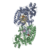
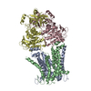
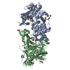
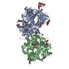
| Title | Teneurin3 monomer | |||||||||
 Components Components | Odz3 protein | |||||||||
 Keywords Keywords | CELL ADHESION / Neuronal cell adhesion / bacterial toxin-like | |||||||||
| Function / homology |  Function and homology information Function and homology informationregulation of homophilic cell adhesion / synaptic membrane adhesion / camera-type eye morphogenesis / homophilic cell-cell adhesion / neuron development / presynaptic active zone membrane / cell adhesion molecule binding / positive regulation of neuron projection development / cell differentiation / neuron projection ...regulation of homophilic cell adhesion / synaptic membrane adhesion / camera-type eye morphogenesis / homophilic cell-cell adhesion / neuron development / presynaptic active zone membrane / cell adhesion molecule binding / positive regulation of neuron projection development / cell differentiation / neuron projection / protein heterodimerization activity / axon / glutamatergic synapse / signal transduction / protein homodimerization activity / membrane / identical protein binding / plasma membrane Similarity search - Function | |||||||||
| Biological species |  | |||||||||
| Method | ELECTRON MICROSCOPY / single particle reconstruction / cryo EM / Resolution: 3.8 Å | |||||||||
 Authors Authors | Janssen, B.J.C. / Meijer, D.H.M. / van Bezouwen, L.S. | |||||||||
| Funding support |  Netherlands, 2items Netherlands, 2items
| |||||||||
 Citation Citation | Journal: Nat Commun / Year: 2018 Title: Structures of Teneurin adhesion receptors reveal an ancient fold for cell-cell interaction. Authors: Verity A Jackson / Dimphna H Meijer / Maria Carrasquero / Laura S van Bezouwen / Edward D Lowe / Colin Kleanthous / Bert J C Janssen / Elena Seiradake /  Abstract: Teneurins are ancient cell-cell adhesion receptors that are vital for brain development and synapse organisation. They originated in early metazoan evolution through a horizontal gene transfer event ...Teneurins are ancient cell-cell adhesion receptors that are vital for brain development and synapse organisation. They originated in early metazoan evolution through a horizontal gene transfer event when a bacterial YD-repeat toxin fused to a eukaryotic receptor. We present X-ray crystallography and cryo-EM structures of two Teneurins, revealing a ~200 kDa extracellular super-fold in which eight sub-domains form an intricate structure centred on a spiralling YD-repeat shell. An alternatively spliced loop, which is implicated in homophilic Teneurin interaction and specificity, is exposed and thus poised for interaction. The N-terminal side of the shell is 'plugged' via a fibronectin-plug domain combination, which defines a new class of YD proteins. Unexpectedly, we find that these proteins are widespread amongst modern bacteria, suggesting early metazoan receptor evolution from a distinct class of proteins, which today includes both bacterial proteins and eukaryotic Teneurins. | |||||||||
| History |
|
- Structure visualization
| Movie |
Movie viewer |
|---|---|
| Structure viewer | Molecule: MolmilJmol/JSmol |
- Downloads & links
-Download
| PDBx/mmCIF format | 6fay.cif.gz | 291.9 KB | Display | PDBx/mmCIF format |
|---|---|---|---|---|
| PDB format | pdb6fay.ent.gz | 221.7 KB | Display | PDB format |
| PDBx/mmJSON format | 6fay.json.gz | Tree view | PDBx/mmJSON format | |
| Others |  Other downloads Other downloads |
-Validation report
| Arichive directory | https://data.pdbj.org/pub/pdb/validation_reports/fa/6fayftp://data.pdbj.org/pub/pdb/validation_reports/fa/6fay | HTTPS FTP |
|---|
-Related structure data
| Related structure data |  4219MC 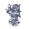 6fb3C M: map data used to model this data C: citing same article ( |
|---|---|
| Similar structure data |


-Links
 PDBj
PDBj


- Assembly
| Deposited unit | 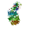
|
|---|---|
| 1 |
|
-Components
| #1: Protein | Mass: 211429.734 Da / Num. of mol.: 1 Source method: isolated from a genetically manipulated source Source: (gene. exp.)  Homo sapiens (human) / References: UniProt: B7ZNJ5, UniProt: Q9WTS6*PLUS Homo sapiens (human) / References: UniProt: B7ZNJ5, UniProt: Q9WTS6*PLUS | ||
|---|---|---|---|
| #2: Sugar | ChemComp-NAG /   Type: D-saccharide, beta linking / Mass: 221.208 Da / Num. of mol.: 5 Type: D-saccharide, beta linking / Mass: 221.208 Da / Num. of mol.: 5Source method: isolated from a genetically manipulated source Formula: C8H15NO6 Has protein modification | Y | |
-Experimental details
-Experiment
| Experiment | Method: ELECTRON MICROSCOPY |
|---|---|
| EM experiment | Aggregation state: PARTICLE / 3D reconstruction method: single particle reconstruction |
- Sample preparation
| Component | Name: Teneurin3 monomer / Type: ORGANELLE OR CELLULAR COMPONENT / Entity ID: #1 / Source: RECOMBINANT | |||||||||||||||
|---|---|---|---|---|---|---|---|---|---|---|---|---|---|---|---|---|
| Molecular weight | Value: 0.22 MDa / Experimental value: NO | |||||||||||||||
| Source (natural) | Organism: | |||||||||||||||
| Source (recombinant) | Organism: Homo sapiens (human) | |||||||||||||||
| Buffer solution | pH: 9 | |||||||||||||||
| Buffer component |
| |||||||||||||||
| Specimen | Conc.: 0.1 mg/ml / Embedding applied: NO / Shadowing applied: NO / Staining applied: NO / Vitrification applied: YES / Details: This sample was monodisperse | |||||||||||||||
| Specimen support | Grid material: COPPER / Grid mesh size: 300 divisions/in. / Grid type: Quantifoil R2/2 | |||||||||||||||
| Vitrification | Instrument: FEI VITROBOT MARK IV / Cryogen name: ETHANE / Humidity: 95 % |
- Electron microscopy imaging
| Experimental equipment |  Model: Talos Arctica / Image courtesy: FEI Company |
|---|---|
| Microscopy | Model: FEI TALOS ARCTICA |
| Electron gun | Electron source:  FIELD EMISSION GUN / Accelerating voltage: 200 kV / Illumination mode: OTHER FIELD EMISSION GUN / Accelerating voltage: 200 kV / Illumination mode: OTHER |
| Electron lens | Mode: OTHER / Nominal magnification: 130000 X / Nominal defocus max: 3000 nm / Nominal defocus min: 1200 nm / Cs: 2.7 mm |
| Specimen holder | Cryogen: NITROGEN |
| Image recording | Electron dose: 56 e/Å2 / Detector mode: SUPER-RESOLUTION / Film or detector model: GATAN K2 SUMMIT (4k x 4k) |
- Processing
| Software | Name: PHENIX / Version: 1.13rc1_2958: / Classification: refinement | ||||||||||||||||||||||||
|---|---|---|---|---|---|---|---|---|---|---|---|---|---|---|---|---|---|---|---|---|---|---|---|---|---|
| EM software |
| ||||||||||||||||||||||||
| CTF correction | Type: PHASE FLIPPING AND AMPLITUDE CORRECTION | ||||||||||||||||||||||||
| Particle selection | Num. of particles selected: 435755 | ||||||||||||||||||||||||
| Symmetry | Point symmetry: C1 (asymmetric) | ||||||||||||||||||||||||
| 3D reconstruction | Resolution: 3.8 Å / Resolution method: FSC 0.143 CUT-OFF / Num. of particles: 287402 / Symmetry type: POINT | ||||||||||||||||||||||||
| Atomic model building | B value: 126 / Protocol: OTHER / Space: REAL | ||||||||||||||||||||||||
| Atomic model building | PDB-ID: 6FB3 Accession code: 6FB3 Details: We have used PDB 6FB3 (same publication) as a template model using Modeller v9.16 followed by manual rebuilding in Coot and real-space refinement using Phenix. Source name: PDB / Type: experimental model | ||||||||||||||||||||||||
| Refine LS restraints |
|
