 Movie
Movie Controller

[English] 日本語
 Yorodumi
- PDB-1lv5: Crystal Structure of the Closed Conformation of Bacillus DNA Poly... -
+ Open data

- Basic information
| Entry | Database: PDB / ID: 1lv5 | ||||||
|---|---|---|---|---|---|---|---|
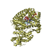
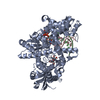
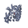
| Title | Crystal Structure of the Closed Conformation of Bacillus DNA Polymerase I Fragment Bound to DNA and dCTP | ||||||
 Components Components |
| ||||||
 Keywords Keywords | TRANSFERASE/DNA / DNA POLYMERASE I / DNA REPLICATION / KLENOW FRAGMENT / PROTEIN-DNA-DNTP COMPLEX / CLOSED CONFORMATION / TRANSFERASE-DNA COMPLEX | ||||||
| Function / homology |  Function and homology information Function and homology information5'-3' exonuclease activity / 3'-5' exonuclease activity / DNA-templated DNA replication / double-strand break repair / DNA-directed DNA polymerase / DNA-directed DNA polymerase activity / DNA binding Similarity search - Function | ||||||
| Biological species |   Geobacillus stearothermophilus (bacteria) Geobacillus stearothermophilus (bacteria) | ||||||
| Method |  X-RAY DIFFRACTION / SYNCHROTRON / MOLECULAR REPLACEMENT / Resolution: 1.95 Å X-RAY DIFFRACTION / SYNCHROTRON / MOLECULAR REPLACEMENT / Resolution: 1.95 Å | ||||||
 Authors Authors | Johnson, S.J. / Taylor, J.S. / Beese, L.S. | ||||||
 Citation Citation | Journal: Proc.Natl.Acad.Sci.USA / Year: 2003 Title: Processive DNA synthesis observed in a polymerase crystal suggests a mechanism for the prevention of frameshift mutations Authors: Johnson, S.J. / Taylor, J.S. / Beese, L.S. #1: Journal: Nature / Year: 1998Title: Visualizing DNA Replication in a Catalytically Active Bacillus DNA Polymerase Crystal Authors: Kiefer, J.R. / Mao, C. / Braman, J.C. / Beese, L.S. #2: Journal: Structure / Year: 1997Title: Crystal Structure of a Thermostable Bacillus DNA Polymerase I Large Fragment at 2.1 A Resolution Authors: Kiefer, J.R. / Mao, C. / Hansen, C.J. / Basehore, S.L. / Hogrefe, H.H. / Braman, J.C. / Beese, L.S. | ||||||
| History |
|
- Structure visualization
| Structure viewer | Molecule: MolmilJmol/JSmol |
|---|
- Downloads & links
-Download
| PDBx/mmCIF format | 1lv5.cif.gz | 273.1 KB | Display | PDBx/mmCIF format |
|---|---|---|---|---|
| PDB format | pdb1lv5.ent.gz | 215.1 KB | Display | PDB format |
| PDBx/mmJSON format | 1lv5.json.gz | Tree view | PDBx/mmJSON format | |
| Others |  Other downloads Other downloads |
-Validation report
| Arichive directory | https://data.pdbj.org/pub/pdb/validation_reports/lv/1lv5ftp://data.pdbj.org/pub/pdb/validation_reports/lv/1lv5 | HTTPS FTP |
|---|
-Related structure data
| Related structure data |  1l3sC  1l3tC  1l3uC  1l3vC  1l5uC  2bdpS C: citing same article ( S: Starting model for refinement |
|---|---|
| Similar structure data |


-Links
 PDBj
PDBj


- Assembly
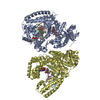
| Deposited unit | 
| ||||||||
|---|---|---|---|---|---|---|---|---|---|
| 1 | 
| ||||||||
| 2 | 
| ||||||||
| Unit cell |
|
-Components
-DNA chain , 2 types, 4 molecules CEDF
| #1: DNA chain | Mass: 3094.043 Da / Num. of mol.: 2 / Source method: obtained synthetically / Details: PRIMER STRAND #2: DNA chain | Mass: 4256.767 Da / Num. of mol.: 2 / Source method: obtained synthetically / Details: Template Strand |
|---|
-Protein , 1 types, 2 molecules AB
| #3: Protein | Mass: 66128.836 Da / Num. of mol.: 2 Fragment: BACILLUS FRAGMENT (ANALOGOUS TO THE E. COLI KLENOW FRAGMENT) Mutation: D329A Source method: isolated from a genetically manipulated source Source: (gene. exp.) Geobacillus stearothermophilus (bacteria)Description: THIS PROTEIN WAS ISOLATED FROM AN AS YET UNNAMED NOVEL STRAIN OF BACILLUS STEAROTHERMOPHILUS (SEE REF 2). Plasmid: pET-30A(+) / Production host: |
|---|
-Non-polymers , 5 types, 193 molecules 


| #4: Chemical |  Mass: 54.938 Da / Num. of mol.: 2 / Source method: obtained synthetically / Formula: Mn Mass: 54.938 Da / Num. of mol.: 2 / Source method: obtained synthetically / Formula: Mn#5: Chemical | ChemComp-MG / |  Mass: 24.305 Da / Num. of mol.: 1 / Source method: obtained synthetically / Formula: Mg Mass: 24.305 Da / Num. of mol.: 1 / Source method: obtained synthetically / Formula: Mg#6: Chemical |  Mass: 96.063 Da / Num. of mol.: 2 / Source method: obtained synthetically / Formula: SO4 Mass: 96.063 Da / Num. of mol.: 2 / Source method: obtained synthetically / Formula: SO4#7: Chemical |  Mass: 467.157 Da / Num. of mol.: 2 / Source method: obtained synthetically / Formula: C9H16N3O13P3 Mass: 467.157 Da / Num. of mol.: 2 / Source method: obtained synthetically / Formula: C9H16N3O13P3#8: Water | ChemComp-HOH / | Mass: 18.015 Da / Num. of mol.: 186 / Source method: isolated from a natural source / Formula: H2O |
|---|
-Experimental details
-Experiment
| Experiment | Method: X-RAY DIFFRACTION / Number of used crystals: 1 |
|---|
- Sample preparation
| Crystal | Density Matthews: 3.12 Å3/Da / Density % sol: 60.63 % | ||||||||||||||||||||||||||||||||||||||||||
|---|---|---|---|---|---|---|---|---|---|---|---|---|---|---|---|---|---|---|---|---|---|---|---|---|---|---|---|---|---|---|---|---|---|---|---|---|---|---|---|---|---|---|---|
| Crystal grow | Temperature: 290 K / Method: vapor diffusion, hanging drop / pH: 5.8 Details: AMMONIUM SULFATE, MAGNESIUM SULFATE, MPD, MES, MANGANESE SULFATE, pH 5.80, VAPOR DIFFUSION, HANGING DROP, temperature 290.0K | ||||||||||||||||||||||||||||||||||||||||||
| Components of the solutions |
| ||||||||||||||||||||||||||||||||||||||||||
| Crystal grow | *PLUS pH: 5.8 | ||||||||||||||||||||||||||||||||||||||||||
| Components of the solutions | *PLUS
|
-Data collection
| Diffraction | Mean temperature: 100 K |
|---|---|
| Diffraction source | Source: SYNCHROTRON / Site: NSLS  / Beamline: X25 / Wavelength: 1.1 Å / Beamline: X25 / Wavelength: 1.1 Å |
| Detector | Type: ADSC QUANTUM 315 / Detector: CCD / Date: Mar 29, 2001 / Details: MIRROR |
| Radiation | Monochromator: Si Crystal / Protocol: SINGLE WAVELENGTH / Monochromatic (M) / Laue (L): M / Scattering type: x-ray |
| Radiation wavelength | Wavelength: 1.1 Å / Relative weight: 1 |
| Reflection | Resolution: 1.95→39.7 Å / Num. all: 129531 / Num. obs: 129531 / % possible obs: 99.8 % / Observed criterion σ(I): -3 / Redundancy: 4.9 % / Biso Wilson estimate: 23.2 Å2 / Rmerge(I) obs: 0.062 / Net I/σ(I): 18.6 |
| Reflection shell | Resolution: 1.95→2.02 Å / Redundancy: 4.1 % / Rmerge(I) obs: 0.48 / Mean I/σ(I) obs: 2.5 / Num. unique all: 12800 / % possible all: 99.3 |
| Reflection | *PLUS Lowest resolution: 50 Å / % possible obs: 74.1 % / Num. measured all: 633019 |
| Reflection shell | *PLUS % possible obs: 39.8 % / Rmerge(I) obs: 0.48 |
- Processing
| Software |
| ||||||||||||||||||||||||||||||||||||
|---|---|---|---|---|---|---|---|---|---|---|---|---|---|---|---|---|---|---|---|---|---|---|---|---|---|---|---|---|---|---|---|---|---|---|---|---|---|
| Refinement | Method to determine structure: MOLECULAR REPLACEMENT Starting model: PDB ENTRY 2BDP Resolution: 1.95→39.7 Å / Rfactor Rfree error: 0.004 / Isotropic thermal model: RESTRAINED / Cross valid method: THROUGHOUT / σ(F): 2 / Stereochemistry target values: Engh & Huber Details: The crystal was twinned, with a partial twinning fraction of 0.485. Detwinning of the data was performed by treating the data as perfectly twinned, using the detwin_perfect.inp routine in ...Details: The crystal was twinned, with a partial twinning fraction of 0.485. Detwinning of the data was performed by treating the data as perfectly twinned, using the detwin_perfect.inp routine in CNS. The Bacillus polymerase was co-crystallized with ddATP, dCTP, and a DNA containing a 9 base pair duplex, a 4 base 5' template overhang, and a single base 3' template overhang. The 3' overhang on the template strand assured that the DNA duplex was not bound backwards by the polymerase during crystallization. ddATP was incorporated onto the 3' terminus of the primer strand. dCTP was not incorporated onto the primer strand, but was paired opposite the next template base. Thus, the resulting structure contained a 10 base pair DNA duplex with a 3' primer terminus and a 3 base 5' template overhang. Electron density was observed for all protein side chains except Lysine 298, which was modelled to the beta carbon. The manganese and magnesium atoms were assigned on the basis of low B-factor, proximity to coordinating residues, and by comparison to a related Klentaq polymerase structure (3KTQ). The data prevent a definitive assignment between MG 205 and water.
| ||||||||||||||||||||||||||||||||||||
| Solvent computation | Solvent model: FLAT MODEL / Bsol: 47.4519 Å2 / ksol: 0.364792 e/Å3 | ||||||||||||||||||||||||||||||||||||
| Displacement parameters | Biso mean: 43.9 Å2
| ||||||||||||||||||||||||||||||||||||
| Refine analyze | Luzzati coordinate error free: 0.32 Å / Luzzati sigma a free: 0.33 Å | ||||||||||||||||||||||||||||||||||||
| Refinement step | Cycle: LAST / Resolution: 1.95→39.7 Å
| ||||||||||||||||||||||||||||||||||||
| Refine LS restraints |
| ||||||||||||||||||||||||||||||||||||
| LS refinement shell | Resolution: 1.95→2.02 Å / Rfactor Rfree error: 0.021 / Total num. of bins used: 10
| ||||||||||||||||||||||||||||||||||||
| Xplor file |
| ||||||||||||||||||||||||||||||||||||
| Refinement | *PLUS Lowest resolution: 50 Å / % reflection Rfree: 5 % | ||||||||||||||||||||||||||||||||||||
| Solvent computation | *PLUS | ||||||||||||||||||||||||||||||||||||
| Displacement parameters | *PLUS | ||||||||||||||||||||||||||||||||||||
| Refine LS restraints | *PLUS
|
