[English] 日本語
Yorodumi
- PDB-6fb3: Teneurin 2 Partial Extracellular Domain -

+
Open data


ID or keywords:

Loading...

-
Basic information

Entry
Database: PDB / ID: 6fb3
TitleTeneurin 2 Partial Extracellular Domain
ComponentsTeneurin-2Teneurin
KeywordsCELL ADHESION / TTR / fibronectin / FN-plug / NHL / YD-repeat / antibiotic-binding / colicin / nuclease / cell surface receptor / latrophilin / glycoprotein
Function / homology
Function and homology information


positive regulation of filopodium assembly / heterophilic cell-cell adhesion via plasma membrane cell adhesion molecules / : / neuron development / cell adhesion molecule binding / filopodium / axon guidance / cell-cell adhesion / PML body / cell-cell junction ...positive regulation of filopodium assembly / heterophilic cell-cell adhesion via plasma membrane cell adhesion molecules / : / neuron development / cell adhesion molecule binding / filopodium / axon guidance / cell-cell adhesion / PML body / cell-cell junction / cell junction / growth cone / postsynaptic membrane / dendritic spine / neuron projection / protein heterodimerization activity / signaling receptor binding / dendrite / synapse / calcium ion binding / Golgi apparatus / negative regulation of transcription by RNA polymerase II / endoplasmic reticulum / protein homodimerization activity / nucleus / plasma membrane
Similarity search - Function
Teneurin intracellular, N-terminal / Teneurin Intracellular Region / Teneurin N-terminal domain profile. / Tox-GHH domain / GHH signature containing HNH/Endo VII superfamily nuclease toxin / YD repeat / Rhs repeat-associated core / Carboxypeptidase-like, regulatory domain superfamily / Six-bladed beta-propeller, TolB-like / Quinoprotein alcohol dehydrogenase-like superfamily ...Teneurin intracellular, N-terminal / Teneurin Intracellular Region / Teneurin N-terminal domain profile. / Tox-GHH domain / GHH signature containing HNH/Endo VII superfamily nuclease toxin / YD repeat / Rhs repeat-associated core / Carboxypeptidase-like, regulatory domain superfamily / Six-bladed beta-propeller, TolB-like / Quinoprotein alcohol dehydrogenase-like superfamily / EGF-like calcium-binding domain / Calcium-binding EGF-like domain / Epidermal growth factor-like domain. / EGF-like domain profile. / EGF-like domain signature 2. / EGF-like domain signature 1. / EGF-like domain
Similarity search - Domain/homology
Biological speciesGallus gallus (chicken)
MethodX-RAY DIFFRACTION / SYNCHROTRON / SAD / Resolution: 2.38 Å
AuthorsJackson, V.A. / Carrasquero, M. / Lowe, E.D. / Seiradake, E.
Funding support United Kingdom, 2items
OrganizationGrant numberCountry
Wellcome Trust202827/Z/16/Z United Kingdom
Medical Research Council (United Kingdom)MR/L018039/1 United Kingdom
Citation
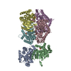
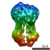
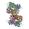
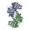
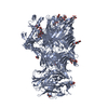
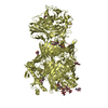
Journal: Nat Commun / Year: 2018
Title: Structures of Teneurin adhesion receptors reveal an ancient fold for cell-cell interaction.
Authors: Verity A Jackson / Dimphna H Meijer / Maria Carrasquero / Laura S van Bezouwen / Edward D Lowe / Colin Kleanthous / Bert J C Janssen / Elena Seiradake /
Abstract: Teneurins are ancient cell-cell adhesion receptors that are vital for brain development and synapse organisation. They originated in early metazoan evolution through a horizontal gene transfer event ...Teneurins are ancient cell-cell adhesion receptors that are vital for brain development and synapse organisation. They originated in early metazoan evolution through a horizontal gene transfer event when a bacterial YD-repeat toxin fused to a eukaryotic receptor. We present X-ray crystallography and cryo-EM structures of two Teneurins, revealing a ~200 kDa extracellular super-fold in which eight sub-domains form an intricate structure centred on a spiralling YD-repeat shell. An alternatively spliced loop, which is implicated in homophilic Teneurin interaction and specificity, is exposed and thus poised for interaction. The N-terminal side of the shell is 'plugged' via a fibronectin-plug domain combination, which defines a new class of YD proteins. Unexpectedly, we find that these proteins are widespread amongst modern bacteria, suggesting early metazoan receptor evolution from a distinct class of proteins, which today includes both bacterial proteins and eukaryotic Teneurins.
History
DepositionDec 18, 2017Deposition site: PDBE / Processing site: PDBE
Revision 1.0Mar 28, 2018Provider: repository / Type: Initial release
Revision 1.1Mar 27, 2019Group: Data collection / Derived calculations / Other
Category: diffrn_source / pdbx_database_status ...diffrn_source / pdbx_database_status / pdbx_struct_assembly / pdbx_struct_assembly_gen / pdbx_struct_assembly_prop
Item: _diffrn_source.pdbx_synchrotron_site / _pdbx_database_status.process_site
Revision 1.2Jul 10, 2019Group: Data collection / Category: diffrn_source / Item: _diffrn_source.pdbx_synchrotron_site
Revision 2.0Jul 29, 2020Group: Atomic model / Data collection ...Atomic model / Data collection / Derived calculations / Structure summary
Category: atom_site / atom_site_anisotrop ...atom_site / atom_site_anisotrop / chem_comp / entity / pdbx_branch_scheme / pdbx_chem_comp_identifier / pdbx_entity_branch / pdbx_entity_branch_descriptor / pdbx_entity_branch_link / pdbx_entity_branch_list / pdbx_entity_nonpoly / pdbx_nonpoly_scheme / pdbx_struct_assembly_gen / struct_asym / struct_conn / struct_site / struct_site_gen
Item: _atom_site.B_iso_or_equiv / _atom_site.Cartn_x ..._atom_site.B_iso_or_equiv / _atom_site.Cartn_x / _atom_site.Cartn_y / _atom_site.Cartn_z / _atom_site.auth_asym_id / _atom_site.auth_atom_id / _atom_site.auth_comp_id / _atom_site.auth_seq_id / _atom_site.label_asym_id / _atom_site.label_atom_id / _atom_site.label_comp_id / _atom_site.label_entity_id / _atom_site.type_symbol / _atom_site_anisotrop.U[1][1] / _atom_site_anisotrop.U[1][2] / _atom_site_anisotrop.U[1][3] / _atom_site_anisotrop.U[2][2] / _atom_site_anisotrop.U[2][3] / _atom_site_anisotrop.U[3][3] / _atom_site_anisotrop.pdbx_auth_asym_id / _atom_site_anisotrop.pdbx_auth_atom_id / _atom_site_anisotrop.pdbx_auth_comp_id / _atom_site_anisotrop.pdbx_auth_seq_id / _atom_site_anisotrop.pdbx_label_asym_id / _atom_site_anisotrop.pdbx_label_atom_id / _atom_site_anisotrop.pdbx_label_comp_id / _atom_site_anisotrop.type_symbol / _chem_comp.name / _chem_comp.type / _entity.formula_weight / _entity.pdbx_description / _entity.pdbx_number_of_molecules / _entity.type / _pdbx_struct_assembly_gen.asym_id_list / _struct_conn.pdbx_dist_value / _struct_conn.pdbx_role / _struct_conn.ptnr1_auth_asym_id / _struct_conn.ptnr1_auth_seq_id / _struct_conn.ptnr1_label_asym_id / _struct_conn.ptnr1_label_atom_id / _struct_conn.ptnr2_auth_asym_id / _struct_conn.ptnr2_auth_seq_id / _struct_conn.ptnr2_label_asym_id
Description: Carbohydrate remediation / Provider: repository / Type: Remediation

-
Structure visualization

Structure viewerMolecule:
MolmilJmol/JSmol

Downloads & links

-
Assembly

Deposited unit
A: Teneurin-2
B: Teneurin-2
C: Teneurin-2
D: Teneurin-2
hetero molecules


Theoretical massNumber of molelcules
Total (without water)853,68048
Polymers835,1794
Non-polymers18,50144
Water3,297183
1
A: Teneurin-2
hetero molecules


Theoretical massNumber of molelcules
Total (without water)213,42012
Polymers208,7951
Non-polymers4,62511
Water181
TypeNameSymmetry operationNumber
identity operation1_555x,y,z1
2
B: Teneurin-2
hetero molecules


Theoretical massNumber of molelcules
Total (without water)213,42012
Polymers208,7951
Non-polymers4,62511
Water181
TypeNameSymmetry operationNumber
identity operation1_555x,y,z1
3
C: Teneurin-2
hetero molecules


Theoretical massNumber of molelcules
Total (without water)213,42012
Polymers208,7951
Non-polymers4,62511
Water181
TypeNameSymmetry operationNumber
identity operation1_555x,y,z1
4
D: Teneurin-2
hetero molecules


Theoretical massNumber of molelcules
Total (without water)213,42012
Polymers208,7951
Non-polymers4,62511
Water181
TypeNameSymmetry operationNumber
identity operation1_555x,y,z1
Unit cell
Length a, b, c (Å)88.560, 452.560, 146.360
Angle α, β, γ (deg.)90.00, 95.12, 90.00
Int Tables number4
Space group name H-MP1211

-
Components

#1: Protein
Teneurin-2 / Teneurin / Ten-2 / Neurestin / Protein Odd Oz/ten-m homolog 2 / Tenascin-M2 / Ten-m2 / Teneurin transmembrane protein 2


Mass: 208794.688 Da / Num. of mol.: 4
Source method: isolated from a genetically manipulated source
Source: (gene. exp.) Gallus gallus (chicken) / Gene: TENM2, ODZ2, TNM2 / Plasmid: pHLSec / Cell (production host): HEK293T / Production host: 
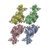
Homo sapiens (human) / References: UniProt: Q9DER5
#2: Polysaccharide
alpha-D-mannopyranose-(1-2)-alpha-D-mannopyranose-(1-2)-alpha-D-mannopyranose-(1-3)-[alpha-D- ...alpha-D-mannopyranose-(1-2)-alpha-D-mannopyranose-(1-2)-alpha-D-mannopyranose-(1-3)-[alpha-D-mannopyranose-(1-3)-alpha-D-mannopyranose-(1-6)]beta-D-mannopyranose-(1-4)-2-acetamido-2-deoxy-beta-D-glucopyranose-(1-4)-2-acetamido-2-deoxy-beta-D-glucopyranose


Type: oligosaccharide / Mass: 1397.245 Da / Num. of mol.: 4
Source method: isolated from a genetically manipulated source
DescriptorTypeProgram
DManpa1-2DManpa1-2DManpa1-3[DManpa1-3DManpa1-6]DManpb1-4DGlcpNAcb1-4DGlcpNAcb1-Glycam Condensed SequenceGMML 1.0
WURCS=2.0/3,8,7/[a2122h-1b_1-5_2*NCC/3=O][a1122h-1b_1-5][a1122h-1a_1-5]/1-1-2-3-3-3-3-3/a4-b1_b4-c1_c3-d1_c6-g1_d2-e1_e2-f1_g3-h1WURCSPDB2Glycan 1.1.0
[]{[(4+1)][b-D-GlcpNAc]{[(4+1)][b-D-GlcpNAc]{[(4+1)][b-D-Manp]{[(3+1)][a-D-Manp]{[(2+1)][a-D-Manp]{[(2+1)][a-D-Manp]{}}}[(6+1)][a-D-Manp]{[(3+1)][a-D-Manp]{}}}}}}LINUCSPDB-CARE
#3: Polysaccharide
2-acetamido-2-deoxy-beta-D-glucopyranose-(1-4)-2-acetamido-2-deoxy-beta-D-glucopyranose


Type: oligosaccharide / Mass: 424.401 Da / Num. of mol.: 20
Source method: isolated from a genetically manipulated source
DescriptorTypeProgram
DGlcpNAcb1-4DGlcpNAcb1-Glycam Condensed SequenceGMML 1.0
WURCS=2.0/1,2,1/[a2122h-1b_1-5_2*NCC/3=O]/1-1/a4-b1WURCSPDB2Glycan 1.1.0
[]{[(4+1)][b-D-GlcpNAc]{[(4+1)][b-D-GlcpNAc]{}}}LINUCSPDB-CARE
#4: Sugar
ChemComp-NAG / 2-acetamido-2-deoxy-beta-D-glucopyranose / N-Acetylglucosamine


Type: D-saccharide, beta linking / Mass: 221.208 Da / Num. of mol.: 20
Source method: isolated from a genetically manipulated source
Formula: C8H15NO6
IdentifierTypeProgram
DGlcpNAcbCONDENSED IUPAC CARBOHYDRATE SYMBOLGMML 1.0
N-acetyl-b-D-glucopyranosamineCOMMON NAMEGMML 1.0
b-D-GlcpNAcIUPAC CARBOHYDRATE SYMBOLPDB-CARE 1.0
GlcNAcSNFG CARBOHYDRATE SYMBOLGMML 1.0
#5: Water ChemComp-HOH / water / Water


Mass: 18.015 Da / Num. of mol.: 183 / Source method: isolated from a natural source / Formula: H2O

-
Experimental details

-
Experiment

ExperimentMethod: X-RAY DIFFRACTION / Number of used crystals: 1

-
Sample preparation

CrystalDensity Matthews: 3.5 Å3/Da / Density % sol: 64.83 %
Crystal growTemperature: 291.15 K / Method: vapor diffusion / pH: 8.5
Details: 0.1 M Tris (base)/Bicine (pH 8.5), 20% v/v glycerol, 10% w/v PEG4K, 30 mM di-ethyleneglycol, 30 mM tri-ethyleneglycol, 30 mM tetraethyleneglycol, and 30 mM pentaethyleneglycol

-
Data collection

DiffractionMean temperature: 100 K
Diffraction sourceSource: SYNCHROTRON / Site: Diamond / Beamline: I03 / Wavelength: 0.976 Å
DetectorType: DECTRIS PILATUS3 6M / Detector: PIXEL / Date: May 6, 2017
RadiationProtocol: SINGLE WAVELENGTH / Monochromatic (M) / Laue (L): M / Scattering type: x-ray
Radiation wavelengthWavelength: 0.976 Å / Relative weight: 1
ReflectionResolution: 2.38→90.51 Å / Num. obs: 439054 / % possible obs: 96.3 % / Redundancy: 5.6 % / Biso Wilson estimate: 54.02 Å2 / CC1/2: 0.99 / Rrim(I) all: 0.25 / Net I/σ(I): 4.69
Reflection shellResolution: 2.38→2.44 Å / Redundancy: 1.8 % / Mean I/σ(I) obs: 0.39 / Num. unique obs: 29248 / CC1/2: 0.315 / % possible all: 86.8

-
Processing

Software
NameVersionClassification
BUSTER2.10.3refinement
XDSdata reduction
XDSdata scaling
PHASERphasing
RefinementMethod to determine structure: SAD / Resolution: 2.38→89.38 Å / Cor.coef. Fo:Fc: 0.933 / Cor.coef. Fo:Fc free: 0.922 / SU R Cruickshank DPI: 0.303 / Cross valid method: THROUGHOUT / σ(F): 0 / SU R Blow DPI: 0.279 / SU Rfree Blow DPI: 0.208 / SU Rfree Cruickshank DPI: 0.218
RfactorNum. reflection% reflectionSelection details
Rfree0.242 21455 4.99 %RANDOM
Rwork0.225 ---
obs0.226 429571 94.3 %-
Displacement parametersBiso mean: 76.85 Å2
Baniso -1Baniso -2Baniso -3
1--5.8554 Å20 Å2-0.9532 Å2
2--8.5928 Å20 Å2
3----2.7374 Å2
Refine analyzeLuzzati coordinate error obs: 0.46 Å
Refinement stepCycle: 1 / Resolution: 2.38→89.38 Å
ProteinNucleic acidLigandSolventTotal
Num. atoms58012 0 1216 183 59411
Refine LS restraints
Refine-IDTypeDev idealNumberRestraint functionWeight
X-RAY DIFFRACTIONt_bond_d0.00760644HARMONIC2
X-RAY DIFFRACTIONt_angle_deg0.9982420HARMONIC2
X-RAY DIFFRACTIONt_dihedral_angle_d21156SINUSOIDAL2
X-RAY DIFFRACTIONt_incorr_chiral_ct
X-RAY DIFFRACTIONt_pseud_angle
X-RAY DIFFRACTIONt_trig_c_planes
X-RAY DIFFRACTIONt_gen_planes10280HARMONIC5
X-RAY DIFFRACTIONt_it60644HARMONIC20
X-RAY DIFFRACTIONt_nbd
X-RAY DIFFRACTIONt_omega_torsion2.35
X-RAY DIFFRACTIONt_other_torsion18.34
X-RAY DIFFRACTIONt_improper_torsion
X-RAY DIFFRACTIONt_chiral_improper_torsion8168SEMIHARMONIC5
X-RAY DIFFRACTIONt_sum_occupancies
X-RAY DIFFRACTIONt_utility_distance
X-RAY DIFFRACTIONt_utility_angle
X-RAY DIFFRACTIONt_utility_torsion
X-RAY DIFFRACTIONt_ideal_dist_contact62202SEMIHARMONIC4
LS refinement shellResolution: 2.38→2.44 Å / Total num. of bins used: 20
RfactorNum. reflection% reflection
Rfree0.3191 1318 4.91 %
Rwork0.3066 25518 -
all0.3073 26836 -
obs--79.73 %
Refinement TLS params.

Method: refined / Refine-ID: X-RAY DIFFRACTION

IDL112)L122)L132)L222)L232)L332)S11 (Å °)S12 (Å °)S13 (Å °)S21 (Å °)S22 (Å °)S23 (Å °)S31 (Å °)S32 (Å °)S33 (Å °)T112)T122)T132)T222)T232)T332)Origin x (Å)Origin y (Å)Origin z (Å)
10.27380.29490.0182.6393-0.24910.48120.04-0.06260.15310.4913-0.09180.1471-0.11330.02230.05180.1384-0.0739-0.1204-0.00040.01210.12-20.8857-61.1685-66.9857
20.3075-0.27920.07021.7374-0.23490.3492-0.03470.0095-0.01940.1796-0.042-0.26740.11460.02020.07670.32090.0933-0.0799-0.0084-0.0025-0.0213-15.09-165.603-54.6256
30.16570.0012-0.06410.4401-0.09820.1424-0.0126-0.0150.09520.02170.02170.022-0.00790.0017-0.0092-0.0063-0.0272-0.1222-0.04250.03840.0206-58.1069-60.9673-139.2753
40.3105-0.530.17172.3412-0.48240.1211-0.1065-0.0406-0.17530.37240.17650.54180-0.0462-0.070.23130.10060.0695-0.01690.01980.0487-51.5647-165.294-127.0105
Refinement TLS group
IDRefine-IDRefine TLS-IDSelection details
1X-RAY DIFFRACTION1{ A|* }
2X-RAY DIFFRACTION2{ B|* }
3X-RAY DIFFRACTION3{ C|* }
4X-RAY DIFFRACTION4{ D|* }

+
About Yorodumi

-
News

-
Feb 9, 2022. New format data for meta-information of EMDB entries

New format data for meta-information of EMDB entries

  • Version 3 of the EMDB header file is now the official format.
  • The previous official version 1.9 will be removed from the archive.

Related info.:EMDB header

External links:wwPDB to switch to version 3 of the EMDB data model

-
Aug 12, 2020. Covid-19 info

Covid-19 info

URL: https://pdbj.org/emnavi/covid19.php

New page: Covid-19 featured information page in EM Navigator.

Related info.:Covid-19 info / Mar 5, 2020. Novel coronavirus structure data

+
Mar 5, 2020. Novel coronavirus structure data

Novel coronavirus structure data

Related info.:Yorodumi Speices / Aug 12, 2020. Covid-19 info

External links:COVID-19 featured content - PDBj / Molecule of the Month (242):Coronavirus Proteases

+
Jan 31, 2019. EMDB accession codes are about to change! (news from PDBe EMDB page)

EMDB accession codes are about to change! (news from PDBe EMDB page)

  • The allocation of 4 digits for EMDB accession codes will soon come to an end. Whilst these codes will remain in use, new EMDB accession codes will include an additional digit and will expand incrementally as the available range of codes is exhausted. The current 4-digit format prefixed with “EMD-” (i.e. EMD-XXXX) will advance to a 5-digit format (i.e. EMD-XXXXX), and so on. It is currently estimated that the 4-digit codes will be depleted around Spring 2019, at which point the 5-digit format will come into force.
  • The EM Navigator/Yorodumi systems omit the EMD- prefix.

Related info.:Q: What is EMD? / ID/Accession-code notation in Yorodumi/EM Navigator

External links:EMDB Accession Codes are Changing Soon! / Contact to PDBj

+
Jul 12, 2017. Major update of PDB

Major update of PDB

  • wwPDB released updated PDB data conforming to the new PDBx/mmCIF dictionary.
  • This is a major update changing the version number from 4 to 5, and with Remediation, in which all the entries are updated.
  • In this update, many items about electron microscopy experimental information are reorganized (e.g. em_software).
  • Now, EM Navigator and Yorodumi are based on the updated data.

External links:wwPDB Remediation / Enriched Model Files Conforming to OneDep Data Standards Now Available in the PDB FTP Archive

-
Yorodumi

Thousand views of thousand structures

  • Yorodumi is a browser for structure data from EMDB, PDB, SASBDB, etc.
  • This page is also the successor to EM Navigator detail page, and also detail information page/front-end page for Omokage search.
  • The word "yorodu" (or yorozu) is an old Japanese word meaning "ten thousand". "mi" (miru) is to see.

Related info.:EMDB / PDB / SASBDB / Comparison of 3 databanks / Yorodumi Search / Aug 31, 2016. New EM Navigator & Yorodumi / Yorodumi Papers / Jmol/JSmol / Function and homology information / Changes in new EM Navigator and Yorodumi

Read more