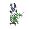
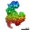
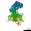
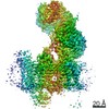
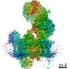
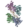
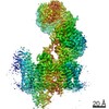
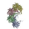
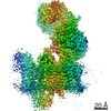
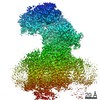
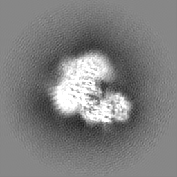
登録情報 データベース : EMDB / ID : EMD-6475タイトル Cryo-EM structure of the rabbit voltage-gated calcium channel Cav1.1 at 4.2 angstrom resolution Reconstruction of a membrane protein 試料 : a membrane protein cryo-sampleタンパク質・ペプチド : Voltage-dependent L-type calcium channel subunit alpha-1Sタンパク質・ペプチド : Voltage-dependent L-type calcium channel subunit beta-2タンパク質・ペプチド : Voltage-dependent calcium channel gamma-1 subunitタンパク質・ペプチド : Voltage-dependent calcium channel subunit alpha-2/delta-1機能・相同性 分子機能 ドメイン・相同性 構成要素
/ / / / / / / / / / / / / / / / / / / / / / / / / / / / / / / / / / / / / / / / / / / / / / / / / / / / / / / / / / / / / / / / / / / / / / / / / / / / / / / / / / / / / / / / / / / / / / / / / / / / / / / / 生物種 Oryctolagus cuniculus (ウサギ)手法 / / 解像度 : 4.2 Å Wu JP / Yan Z / Li ZQ / Yan N ジャーナル : Science / 年 : 2015タイトル : Structure of the voltage-gated calcium channel Cav1.1 complex.著者 : Jianping Wu / Zhen Yan / Zhangqiang Li / Chuangye Yan / Shan Lu / Mengqiu Dong / Nieng Yan / 要旨 : The voltage-gated calcium channel Ca(v)1.1 is engaged in the excitation-contraction coupling of skeletal muscles. The Ca(v)1.1 complex consists of the pore-forming subunit α1 and auxiliary subunits ... The voltage-gated calcium channel Ca(v)1.1 is engaged in the excitation-contraction coupling of skeletal muscles. The Ca(v)1.1 complex consists of the pore-forming subunit α1 and auxiliary subunits α2δ, β, and γ. We report the structure of the rabbit Ca(v)1.1 complex determined by single-particle cryo-electron microscopy. The four homologous repeats of the α1 subunit are arranged clockwise in the extracellular view. The γ subunit, whose structure resembles claudins, interacts with the voltage-sensing domain of repeat IV (VSD(IV)), whereas the cytosolic β subunit is located adjacent to VSD(II) of α1. The α2 subunit interacts with the extracellular loops of repeats I to III through its VWA and Cache1 domains. The structure reveals the architecture of a prototypical eukaryotic Ca(v) channel and provides a framework for understanding the function and disease mechanisms of Ca(v) and Na(v) channels. 履歴 登録 2015年9月28日 - ヘッダ(付随情報) 公開 2015年12月9日 - マップ公開 2015年12月30日 - 更新 2016年1月13日 - 現状 2016年1月13日 処理サイト : PDBj / 状態 : 公開
すべて表示 表示を減らす
 ムービー
ムービー コントローラー


 データを開く
 基本情報
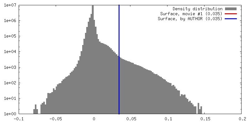
基本情報 マップデータ
 試料
試料 機能・相同性情報
 データ登録者
 引用
引用
 構造の表示
構造の表示
 ダウンロードとリンク
 400_6475.gif
 80_6475.gif
 http://ftp.pdbj.org/pub/emdb/structures/EMD-6475


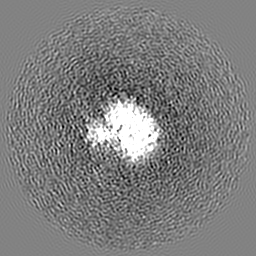
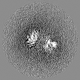
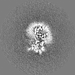
 Z (Sec.)
 Y (Row.)
 X (Col.)


 試料の構成要素
 解析
解析 電子顕微鏡法
 FIELD EMISSION GUN