Movie
Movie Controller

[English] 日本語
 Yorodumi
- EMDB-5514: Icosahedral reconstruction of empty coxsackievirus A9 capsid-inte... -
+ Open data

- Basic information
| Entry | Database: EMDB / ID: EMD-5514 | |||||||||
|---|---|---|---|---|---|---|---|---|---|---|
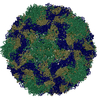
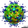
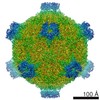
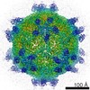
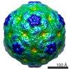
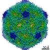
| Title | Icosahedral reconstruction of empty coxsackievirus A9 capsid-integrin alpha v beta 6 complex | |||||||||
 Map data Map data | Icosahedral reconstruction of empty coxsackievirus A9 capsid-integrin alpha v beta 6 complex | |||||||||
 Sample Sample |
| |||||||||
 Keywords Keywords | CVA9-integrin / picornavirus / enterovirus / coxsackievirus A9 / integrin / empty capsid | |||||||||
| Function / homology |  Function and homology information Function and homology informationsymbiont-mediated suppression of host cytoplasmic pattern recognition receptor signaling pathway via inhibition of RIG-I activity / picornain 2A / symbiont-mediated suppression of host mRNA export from nucleus / symbiont genome entry into host cell via pore formation in plasma membrane / picornain 3C / T=pseudo3 icosahedral viral capsid / host cell cytoplasmic vesicle membrane / ribonucleoside triphosphate phosphatase activity / nucleoside-triphosphate phosphatase / channel activity ...symbiont-mediated suppression of host cytoplasmic pattern recognition receptor signaling pathway via inhibition of RIG-I activity / picornain 2A / symbiont-mediated suppression of host mRNA export from nucleus / symbiont genome entry into host cell via pore formation in plasma membrane / picornain 3C / T=pseudo3 icosahedral viral capsid / host cell cytoplasmic vesicle membrane / ribonucleoside triphosphate phosphatase activity / nucleoside-triphosphate phosphatase / channel activity / monoatomic ion transmembrane transport / DNA replication / RNA helicase activity / endocytosis involved in viral entry into host cell / symbiont-mediated activation of host autophagy / RNA-directed RNA polymerase / cysteine-type endopeptidase activity / viral RNA genome replication / RNA-directed RNA polymerase activity / virion attachment to host cell / host cell nucleus / DNA-templated transcription / structural molecule activity / proteolysis / RNA binding / zinc ion binding / ATP binding Similarity search - Function | |||||||||
| Biological species |  Human coxsackievirus A9 Human coxsackievirus A9 | |||||||||
| Method | single particle reconstruction / cryo EM / Resolution: 9.54 Å | |||||||||
 Authors Authors | Shakeel S / Seitsonen JJT / Kajander T / Laurinmaki P / Hyypia T / Susi P / Butcher SJ | |||||||||
 Citation Citation | Journal: J Virol / Year: 2013 Title: Structural and functional analysis of coxsackievirus A9 integrin αvβ6 binding and uncoating. Authors: Shabih Shakeel / Jani J T Seitsonen / Tommi Kajander / Pasi Laurinmäki / Timo Hyypiä / Petri Susi / Sarah J Butcher /  Abstract: Coxsackievirus A9 (CVA9) is an important pathogen of the Picornaviridae family. It utilizes cellular receptors from the integrin αv family for binding to its host cells prior to entry and genome ...Coxsackievirus A9 (CVA9) is an important pathogen of the Picornaviridae family. It utilizes cellular receptors from the integrin αv family for binding to its host cells prior to entry and genome release. Among the integrins tested, it has the highest affinity for αvβ6, which recognizes the arginine-glycine-aspartic acid (RGD) loop present on the C terminus of viral capsid protein, VP1. As the atomic model of CVA9 lacks the RGD loop, we used surface plasmon resonance, electron cryo-microscopy, and image reconstruction to characterize the capsid-integrin interactions and the conformational changes on genome release. We show that the integrin binds to the capsid with nanomolar affinity and that the binding of integrin to the virion does not induce uncoating, thereby implying that further steps are required for release of the genome. Electron cryo-tomography and single-particle image reconstruction revealed variation in the number and conformation of the integrins bound to the capsid, with the integrin footprint mapping close to the predicted site for the exposed RGD loop on VP1. Comparison of empty and RNA-filled capsid reconstructions showed that the capsid undergoes conformational changes when the genome is released, so that the RNA-capsid interactions in the N termini of VP1 and VP4 are lost, VP4 is removed, and the capsid becomes more porous, as has been reported for poliovirus 1, human rhinovirus 2, enterovirus 71, and coxsackievirus A7. These results are important for understanding the structural basis of integrin binding to CVA9 and the molecular events leading to CVA9 cell entry and uncoating. | |||||||||
| History |
|
- Structure visualization
| Movie |
Movie viewer |
|---|---|
| Structure viewer | EM map: SurfViewMolmilJmol/JSmol |
| Supplemental images |

- Downloads & links
-EMDB archive
| Map data | emd_5514.map.gz | 3.6 MB | EMDB map data format | |
|---|---|---|---|---|
| Header (meta data) | emd-5514-v30.xmlemd-5514.xml | 9.6 KB 9.6 KB | Display Display | EMDB header |
| Images | 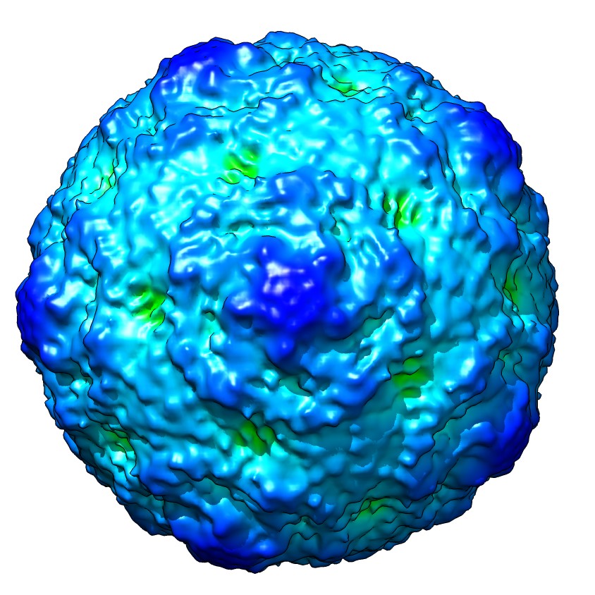 emd_5514_1.jpg emd_5514_1.jpg | 162.3 KB | ||
| Archive directory |  http://ftp.pdbj.org/pub/emdb/structures/EMD-5514ftp://ftp.pdbj.org/pub/emdb/structures/EMD-5514 http://ftp.pdbj.org/pub/emdb/structures/EMD-5514ftp://ftp.pdbj.org/pub/emdb/structures/EMD-5514 | HTTPS FTP |
-Related structure data
| Related structure data |  3j2jMC 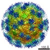 5512C 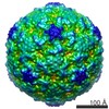 5515C 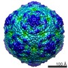 5516C 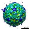 5517C 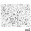 5519C M: atomic model generated by this map C: citing same article ( |
|---|---|
| Similar structure data |
-Links
| EMDB pages | EMDB (EBI/PDBe) / EMDataResource |
|---|---|
| Related items in Molecule of the Month |


-Map
| File | Download / File: emd_5514.map.gz / Format: CCP4 / Size: 15.1 MB / Type: IMAGE STORED AS SIGNED INTEGER (2 BYTES) | ||||||||||||||||||||||||||||||||||||||||||||||||||||||||||||||||||||
|---|---|---|---|---|---|---|---|---|---|---|---|---|---|---|---|---|---|---|---|---|---|---|---|---|---|---|---|---|---|---|---|---|---|---|---|---|---|---|---|---|---|---|---|---|---|---|---|---|---|---|---|---|---|---|---|---|---|---|---|---|---|---|---|---|---|---|---|---|---|
| Annotation | Icosahedral reconstruction of empty coxsackievirus A9 capsid-integrin alpha v beta 6 complex | ||||||||||||||||||||||||||||||||||||||||||||||||||||||||||||||||||||
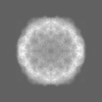
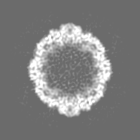
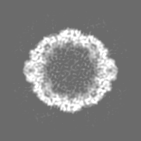
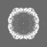
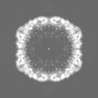
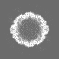
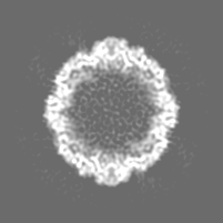
| Projections & slices | Image control
Images are generated by Spider. | ||||||||||||||||||||||||||||||||||||||||||||||||||||||||||||||||||||
| Voxel size | X=Y=Z: 2.26 Å | ||||||||||||||||||||||||||||||||||||||||||||||||||||||||||||||||||||
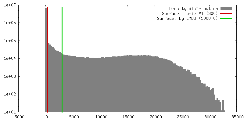
| Density |
| ||||||||||||||||||||||||||||||||||||||||||||||||||||||||||||||||||||
| Symmetry | Space group: 1 | ||||||||||||||||||||||||||||||||||||||||||||||||||||||||||||||||||||
| Details | EMDB XML:
CCP4 map header:
| ||||||||||||||||||||||||||||||||||||||||||||||||||||||||||||||||||||
 Z (Sec.)
 Y (Row.)
 X (Col.)


-Supplemental data
- Sample components
-Entire : empty coxsackievirus A9 capsid in complex with integrin alpha v beta 6
| Entire | Name: empty coxsackievirus A9 capsid in complex with integrin alpha v beta 6 |
|---|---|
| Components |
|
-Supramolecule #1000: empty coxsackievirus A9 capsid in complex with integrin alpha v beta 6
| Supramolecule | Name: empty coxsackievirus A9 capsid in complex with integrin alpha v beta 6 type: sample / ID: 1000 / Number unique components: 2 |
|---|
-Supramolecule #1: Human coxsackievirus A9
| Supramolecule | Name: Human coxsackievirus A9 / type: virus / ID: 1 / Name.synonym: CVA9 / NCBI-ID: 12067 / Sci species name: Human coxsackievirus A9 / Database: NCBI / Virus type: VIRION / Virus isolate: SPECIES / Virus enveloped: No / Virus empty: Yes / Syn species name: CVA9 |
|---|---|
| Host (natural) | Organism:  Homo sapiens (human) / synonym: VERTEBRATES Homo sapiens (human) / synonym: VERTEBRATES |
| Virus shell | Shell ID: 1 / T number (triangulation number): 1 |
-Experimental details
-Structure determination
| Method | cryo EM |
|---|---|
 Processing Processing | single particle reconstruction |
| Aggregation state | particle |
-Sample preparation
| Vitrification | Cryogen name: ETHANE / Instrument: GATAN CRYOPLUNGE 3 / Method: manual plunging |
|---|
- Electron microscopy
| Microscope | FEI TECNAI F20 |
|---|---|
| Date | Feb 1, 2011 |
| Image recording | Digitization - Scanner: OTHER / Digitization - Sampling interval: 7 µm / Number real images: 147 |
| Electron beam | Acceleration voltage: 200 kV / Electron source:  FIELD EMISSION GUN FIELD EMISSION GUN |
| Electron optics | Calibrated magnification: 62000 / Illumination mode: FLOOD BEAM / Imaging mode: BRIGHT FIELD / Nominal defocus max: 4.12 µm / Nominal defocus min: 0.83 µm / Nominal magnification: 62000 |
| Sample stage | Specimen holder: GATAN 626 / Specimen holder model: GATAN LIQUID NITROGEN |
| Experimental equipment |  Model: Tecnai F20 / Image courtesy: FEI Company |
-Image processing
| CTF correction | Details: whole micrograph |
|---|---|
| Final reconstruction | Algorithm: OTHER / Resolution.type: BY AUTHOR / Resolution: 9.54 Å / Resolution method: FSC 0.5 CUT-OFF / Software - Name: AUTO3DEM / Number images used: 1200 |