Movie
Movie Controller


+ Open data

- Basic information
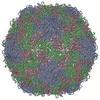
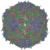
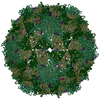
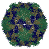
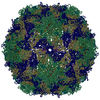
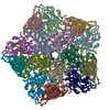
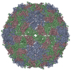
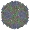
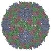
| Entry | Database: PDB / ID: 3j2j | ||||||
|---|---|---|---|---|---|---|---|
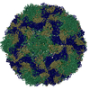
| Title | Empty coxsackievirus A9 capsid | ||||||
 Components Components |
| ||||||
 Keywords Keywords | VIRUS / CVA9-integrin / picornavirus / enterovirus / empty capsid | ||||||
| Function / homology |  Function and homology information Function and homology informationsymbiont-mediated suppression of host cytoplasmic pattern recognition receptor signaling pathway via inhibition of RIG-I activity / picornain 2A / symbiont-mediated suppression of host mRNA export from nucleus / symbiont genome entry into host cell via pore formation in plasma membrane / picornain 3C / T=pseudo3 icosahedral viral capsid / host cell cytoplasmic vesicle membrane / ribonucleoside triphosphate phosphatase activity / nucleoside-triphosphate phosphatase / channel activity ...symbiont-mediated suppression of host cytoplasmic pattern recognition receptor signaling pathway via inhibition of RIG-I activity / picornain 2A / symbiont-mediated suppression of host mRNA export from nucleus / symbiont genome entry into host cell via pore formation in plasma membrane / picornain 3C / T=pseudo3 icosahedral viral capsid / host cell cytoplasmic vesicle membrane / ribonucleoside triphosphate phosphatase activity / nucleoside-triphosphate phosphatase / channel activity / monoatomic ion transmembrane transport / DNA replication / RNA helicase activity / endocytosis involved in viral entry into host cell / symbiont-mediated activation of host autophagy / RNA-directed RNA polymerase / cysteine-type endopeptidase activity / viral RNA genome replication / RNA-directed RNA polymerase activity / virion attachment to host cell / host cell nucleus / DNA-templated transcription / structural molecule activity / proteolysis / RNA binding / zinc ion binding / ATP binding Similarity search - Function | ||||||
| Biological species |  Human coxsackievirus A9 Human coxsackievirus A9 | ||||||
| Method | ELECTRON MICROSCOPY / single particle reconstruction / cryo EM / Resolution: 9.54 Å | ||||||
 Authors Authors | Shakeel, S. / Seitsonen, J.J.T. / Kajander, T. / Laurinmaki, P. / Hyypia, T. / Susi, P. / Butcher, S.J. | ||||||
 Citation Citation | Journal: J Virol / Year: 2013 Title: Structural and functional analysis of coxsackievirus A9 integrin αvβ6 binding and uncoating. Authors: Shabih Shakeel / Jani J T Seitsonen / Tommi Kajander / Pasi Laurinmäki / Timo Hyypiä / Petri Susi / Sarah J Butcher /  Abstract: Coxsackievirus A9 (CVA9) is an important pathogen of the Picornaviridae family. It utilizes cellular receptors from the integrin αv family for binding to its host cells prior to entry and genome ...Coxsackievirus A9 (CVA9) is an important pathogen of the Picornaviridae family. It utilizes cellular receptors from the integrin αv family for binding to its host cells prior to entry and genome release. Among the integrins tested, it has the highest affinity for αvβ6, which recognizes the arginine-glycine-aspartic acid (RGD) loop present on the C terminus of viral capsid protein, VP1. As the atomic model of CVA9 lacks the RGD loop, we used surface plasmon resonance, electron cryo-microscopy, and image reconstruction to characterize the capsid-integrin interactions and the conformational changes on genome release. We show that the integrin binds to the capsid with nanomolar affinity and that the binding of integrin to the virion does not induce uncoating, thereby implying that further steps are required for release of the genome. Electron cryo-tomography and single-particle image reconstruction revealed variation in the number and conformation of the integrins bound to the capsid, with the integrin footprint mapping close to the predicted site for the exposed RGD loop on VP1. Comparison of empty and RNA-filled capsid reconstructions showed that the capsid undergoes conformational changes when the genome is released, so that the RNA-capsid interactions in the N termini of VP1 and VP4 are lost, VP4 is removed, and the capsid becomes more porous, as has been reported for poliovirus 1, human rhinovirus 2, enterovirus 71, and coxsackievirus A7. These results are important for understanding the structural basis of integrin binding to CVA9 and the molecular events leading to CVA9 cell entry and uncoating. | ||||||
| History |
|
- Structure visualization
| Movie |
Movie viewer |
|---|---|
| Structure viewer | Molecule: MolmilJmol/JSmol |
- Downloads & links
-Download
| PDBx/mmCIF format | 3j2j.cif.gz | 134.9 KB | Display | PDBx/mmCIF format |
|---|---|---|---|---|
| PDB format | pdb3j2j.ent.gz | 100.8 KB | Display | PDB format |
| PDBx/mmJSON format | 3j2j.json.gz | Tree view | PDBx/mmJSON format | |
| Others |  Other downloads Other downloads |
-Validation report
| Arichive directory | https://data.pdbj.org/pub/pdb/validation_reports/j2/3j2jftp://data.pdbj.org/pub/pdb/validation_reports/j2/3j2j | HTTPS FTP |
|---|
-Related structure data
| Related structure data | 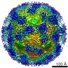 5514MC 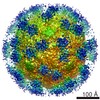 5512C  5515C 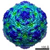 5516C 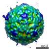 5517C 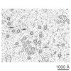 5519C M: map data used to model this data C: citing same article ( |
|---|---|
| Similar structure data |
-Links
 PDBj
PDBj

- Assembly
| Deposited unit | 
|
|---|---|
| 1 | x 60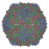
|
| 2 |
|
| 3 | x 5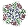
|
| 4 | x 6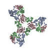
|
| 5 | 
|
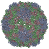
| Symmetry | Point symmetry: (Schoenflies symbol: I (icosahedral)) |
-Components
| #1: Protein | Mass: 25541.980 Da / Num. of mol.: 1 / Fragment: UNP residues 631-852 / Source method: isolated from a natural source / Source: (natural) Human coxsackievirus A9 / References: UniProt: P21404 |
|---|---|
| #2: Protein | Mass: 26335.072 Da / Num. of mol.: 1 / Fragment: UNP residues 331-568 / Source method: isolated from a natural source / Source: (natural) Human coxsackievirus A9 / References: UniProt: P21404 |
| #3: Protein | Mass: 27919.494 Da / Num. of mol.: 1 / Fragment: UNP residues 79-330 / Source method: isolated from a natural source / Source: (natural) Human coxsackievirus A9 / References: UniProt: P21404 |
-Experimental details
-Experiment
| Experiment | Method: ELECTRON MICROSCOPY |
|---|---|
| EM experiment | Aggregation state: PARTICLE / 3D reconstruction method: single particle reconstruction |
- Sample preparation
| Component | Name: Empty coxsackievirus A9 capsid in complex with integrin alpha v beta 6 Type: COMPLEX / Synonym: CVA9 |
|---|---|
| Details of virus | Empty: YES / Enveloped: NO / Host category: VERTEBRATES / Isolate: SPECIES / Type: VIRION |
| Natural host | Organism: Homo sapiens |
| Specimen | Embedding applied: NO / Shadowing applied: NO / Staining applied: NO / Vitrification applied: YES |
| Vitrification | Instrument: GATAN CRYOPLUNGE 3 / Cryogen name: ETHANE Details: manual plunging into liquid ethane (GATAN CRYOPLUNGE 3) Method: manual plunging |
- Electron microscopy imaging
| Experimental equipment |  Model: Tecnai F20 / Image courtesy: FEI Company |
|---|---|
| Microscopy | Model: FEI TECNAI F20 / Date: Feb 1, 2011 |
| Electron gun | Electron source:  FIELD EMISSION GUN / Accelerating voltage: 200 kV / Illumination mode: FLOOD BEAM FIELD EMISSION GUN / Accelerating voltage: 200 kV / Illumination mode: FLOOD BEAM |
| Electron lens | Mode: BRIGHT FIELD / Nominal magnification: 62000 X / Calibrated magnification: 62000 X / Nominal defocus max: 4120 nm / Nominal defocus min: 830 nm / Camera length: 0 mm |
| Specimen holder | Specimen holder model: GATAN LIQUID NITROGEN / Specimen holder type: GATAN 626 / Tilt angle max: 0 ° / Tilt angle min: 0 ° |
| Radiation | Protocol: SINGLE WAVELENGTH / Monochromatic (M) / Laue (L): M / Scattering type: x-ray |
| Radiation wavelength | Relative weight: 1 |
- Processing
| EM software |
| ||||||||||||
|---|---|---|---|---|---|---|---|---|---|---|---|---|---|
| CTF correction | Details: whole micrograph | ||||||||||||
| Symmetry | Point symmetry: I (icosahedral) | ||||||||||||
| 3D reconstruction | Method: polar Fourier transform / Resolution: 9.54 Å / Resolution method: FSC 0.5 CUT-OFF / Num. of particles: 1200 / Nominal pixel size: 2.26 Å / Actual pixel size: 2.26 Å / Symmetry type: POINT | ||||||||||||
| Atomic model building | Protocol: FLEXIBLE FIT / Space: REAL / Details: REFINEMENT PROTOCOL--flexible fitting | ||||||||||||
| Atomic model building | 3D fitting-ID: 1 / Accession code: 1D4M / Initial refinement model-ID: 1 / PDB-ID: 1D4M / Source name: PDB / Type: experimental model
| ||||||||||||
| Refinement step | Cycle: LAST
|