 Movie
Movie Controller

[English] 日本語
 Yorodumi
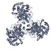
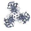
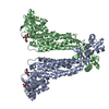
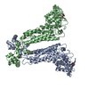
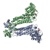
- PDB-4dya: Crystal Structure of WSN/A Influenza Nucleoprotein with BMS-88598... -
+ Open data

- Basic information
| Entry | Database: PDB / ID: 4dya | ||||||
|---|---|---|---|---|---|---|---|
| Title | Crystal Structure of WSN/A Influenza Nucleoprotein with BMS-885986 Ligand Bound | ||||||
 Components Components | Nucleocapsid protein | ||||||
 Keywords Keywords | VIRAL PROTEIN | ||||||
| Function / homology |  Function and homology information Function and homology informationhelical viral capsid / viral penetration into host nucleus / host cell / viral nucleocapsid / ribonucleoprotein complex / symbiont entry into host cell / host cell nucleus / structural molecule activity / RNA binding / identical protein binding Similarity search - Function | ||||||
| Biological species |   Influenza A virus Influenza A virus | ||||||
| Method |  X-RAY DIFFRACTION / SYNCHROTRON / MOLECULAR REPLACEMENT / Resolution: 2.75 Å X-RAY DIFFRACTION / SYNCHROTRON / MOLECULAR REPLACEMENT / Resolution: 2.75 Å | ||||||
 Authors Authors | Lewis, H.A. / Baldwin, E.T. / Steinbacher, S. / Maskos, K. / Mortl, M. / Kiefersauer, R. / Edavettal, S. / McDonnell, P.A. / Pearce, B.C. / Langley, D.R. | ||||||
 Citation Citation | Journal: To be Published Title: To be determined Authors: Lewis, H.A. / Baldwin, E.T. / Steinbacher, S. / Maskos, K. / Mortl, M. / Kiefersauer, R. / Edavettal, S. / McDonnell, P.A. / Pearce, B.C. / Langley, D.R. | ||||||
| History |
|
- Structure visualization
| Structure viewer | Molecule: MolmilJmol/JSmol |
|---|
- Downloads & links
-Download
| PDBx/mmCIF format | 4dya.cif.gz | 170.3 KB | Display | PDBx/mmCIF format |
|---|---|---|---|---|
| PDB format | pdb4dya.ent.gz | 130.5 KB | Display | PDB format |
| PDBx/mmJSON format | 4dya.json.gz | Tree view | PDBx/mmJSON format | |
| Others |  Other downloads Other downloads |
-Validation report
| Arichive directory | https://data.pdbj.org/pub/pdb/validation_reports/dy/4dyaftp://data.pdbj.org/pub/pdb/validation_reports/dy/4dya | HTTPS FTP |
|---|
-Related structure data
| Related structure data | 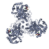 4dybC  4dynC 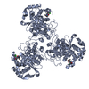 4dypC 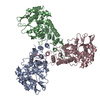 4dysC 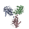 4dytC  5iboC  6dqcC  6dqdC  6dqeC  6dqfC  7rt0C  9bqnC C: citing same article ( |
|---|---|
| Similar structure data |


-Links
 PDBj
PDBj- Assembly
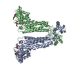
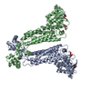
| Deposited unit | 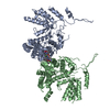
| ||||||||
|---|---|---|---|---|---|---|---|---|---|
| 1 | 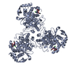
| ||||||||
| 2 | 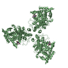
| ||||||||
| Unit cell |
|
-Components
| #1: Protein | Mass: 56806.047 Da / Num. of mol.: 2 Source method: isolated from a genetically manipulated source Source: (gene. exp.) Influenza A virus / Strain: A/WSN/1933(H1N1) / Gene: NP / Plasmid: pET30 / Production host:  #2: Chemical | 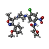  Mass: 565.962 Da / Num. of mol.: 2 / Source method: obtained synthetically / Formula: C27H24ClN5O7 Mass: 565.962 Da / Num. of mol.: 2 / Source method: obtained synthetically / Formula: C27H24ClN5O7#3: Water | ChemComp-HOH / |  Mass: 18.015 Da / Num. of mol.: 24 / Source method: isolated from a natural source / Formula: H2O Mass: 18.015 Da / Num. of mol.: 24 / Source method: isolated from a natural source / Formula: H2O |
|---|
-Experimental details
-Experiment
| Experiment | Method: X-RAY DIFFRACTION / Number of used crystals: 1 |
|---|
- Sample preparation
| Crystal | Density Matthews: 2.26 Å3/Da / Density % sol: 45.55 % |
|---|
-Data collection
| Diffraction source | Source: SYNCHROTRON / Site: SLS  / Beamline: X06SA / Beamline: X06SA | |||||||||||||||||||||||||||||||||||||||||||||||||||||||||||||||||||||||||||||
|---|---|---|---|---|---|---|---|---|---|---|---|---|---|---|---|---|---|---|---|---|---|---|---|---|---|---|---|---|---|---|---|---|---|---|---|---|---|---|---|---|---|---|---|---|---|---|---|---|---|---|---|---|---|---|---|---|---|---|---|---|---|---|---|---|---|---|---|---|---|---|---|---|---|---|---|---|---|---|
| Detector | Type: PSI PILATUS 6M / Detector: PIXEL | |||||||||||||||||||||||||||||||||||||||||||||||||||||||||||||||||||||||||||||
| Radiation | Monochromator: double crystal Si(111) / Protocol: SINGLE WAVELENGTH / Monochromatic (M) / Laue (L): M / Scattering type: x-ray | |||||||||||||||||||||||||||||||||||||||||||||||||||||||||||||||||||||||||||||
| Radiation wavelength | Relative weight: 1 | |||||||||||||||||||||||||||||||||||||||||||||||||||||||||||||||||||||||||||||
| Reflection | Resolution: 2.75→102.881 Å / Num. obs: 26867 / % possible obs: 99.7 % / Observed criterion σ(I): -3 / Biso Wilson estimate: 53.888 Å2 / Rmerge(I) obs: 0.103 / Net I/σ(I): 15.61 | |||||||||||||||||||||||||||||||||||||||||||||||||||||||||||||||||||||||||||||
| Reflection shell | Diffraction-ID: 1
|
- Processing
| Software |
| ||||||||||||||||||||||||||||||||||||||||||||||||||||||||||||||||||||||||||||||||||||||||||
|---|---|---|---|---|---|---|---|---|---|---|---|---|---|---|---|---|---|---|---|---|---|---|---|---|---|---|---|---|---|---|---|---|---|---|---|---|---|---|---|---|---|---|---|---|---|---|---|---|---|---|---|---|---|---|---|---|---|---|---|---|---|---|---|---|---|---|---|---|---|---|---|---|---|---|---|---|---|---|---|---|---|---|---|---|---|---|---|---|---|---|---|
| Refinement | Method to determine structure: MOLECULAR REPLACEMENT / Resolution: 2.75→48.51 Å / Cor.coef. Fo:Fc: 0.902 / Cor.coef. Fo:Fc free: 0.874 / WRfactor Rfree: 0.2517 / WRfactor Rwork: 0.2253 / Occupancy max: 1 / Occupancy min: 1 / FOM work R set: 0.7709 / SU B: 16.124 / SU ML: 0.325 / SU R Cruickshank DPI: 2.0911 / SU Rfree: 0.4094 / Cross valid method: THROUGHOUT / σ(F): 0 / ESU R: 2.091 / ESU R Free: 0.409 / Stereochemistry target values: MAXIMUM LIKELIHOOD
| ||||||||||||||||||||||||||||||||||||||||||||||||||||||||||||||||||||||||||||||||||||||||||
| Solvent computation | Ion probe radii: 0.8 Å / Shrinkage radii: 0.8 Å / VDW probe radii: 1.2 Å / Solvent model: MASK | ||||||||||||||||||||||||||||||||||||||||||||||||||||||||||||||||||||||||||||||||||||||||||
| Displacement parameters | Biso max: 84.4 Å2 / Biso mean: 48.7115 Å2 / Biso min: 7.41 Å2 | ||||||||||||||||||||||||||||||||||||||||||||||||||||||||||||||||||||||||||||||||||||||||||
| Refinement step | Cycle: LAST / Resolution: 2.75→48.51 Å
| ||||||||||||||||||||||||||||||||||||||||||||||||||||||||||||||||||||||||||||||||||||||||||
| Refine LS restraints |
| ||||||||||||||||||||||||||||||||||||||||||||||||||||||||||||||||||||||||||||||||||||||||||
| LS refinement shell | Resolution: 2.75→2.822 Å / Total num. of bins used: 20
|
