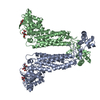
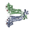
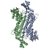
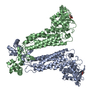
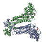
 Movie
Movie Controller


+ Open data

- Basic information
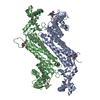
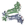
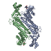
| Entry | Database: PDB / ID: 1kq7 | ||||||
|---|---|---|---|---|---|---|---|
| Title | E315Q Mutant Form of Fumarase C from E.coli | ||||||
 Components Components | FUMARATE HYDRATASE CLASS II | ||||||
 Keywords Keywords | LYASE / fumarate lyase | ||||||
| Function / homology |  Function and homology information Function and homology informationfumarate hydratase activity / fumarate hydratase / fumarate metabolic process / malate metabolic process / tricarboxylic acid cycle / response to oxidative stress / identical protein binding / cytoplasm Similarity search - Function | ||||||
| Biological species |  | ||||||
| Method |  X-RAY DIFFRACTION / MOLECULAR REPLACEMENT / Resolution: 2.6 Å X-RAY DIFFRACTION / MOLECULAR REPLACEMENT / Resolution: 2.6 Å | ||||||
 Authors Authors | Weaver, T.M. / Estevez, M. / Skarda, J. / Spencer, J. | ||||||
 Citation Citation | Journal: Protein Sci. / Year: 2002 Title: X-ray crystallographic and kinetic correlation of a clinically observed human fumarase mutation. Authors: Estevez, M. / Skarda, J. / Spencer, J. / Banaszak, L. / Weaver, T.M. | ||||||
| History |
|
- Structure visualization
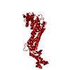
| Structure viewer | Molecule: MolmilJmol/JSmol |
|---|
- Downloads & links
-Download
| PDBx/mmCIF format | 1kq7.cif.gz | 176.8 KB | Display | PDBx/mmCIF format |
|---|---|---|---|---|
| PDB format | pdb1kq7.ent.gz | 141.7 KB | Display | PDB format |
| PDBx/mmJSON format | 1kq7.json.gz | Tree view | PDBx/mmJSON format | |
| Others |  Other downloads Other downloads |
-Validation report
| Arichive directory | https://data.pdbj.org/pub/pdb/validation_reports/kq/1kq7ftp://data.pdbj.org/pub/pdb/validation_reports/kq/1kq7 | HTTPS FTP |
|---|
-Related structure data
| Related structure data |  1fuoS S: Starting model for refinement |
|---|---|
| Similar structure data |


-Links
 PDBj
PDBj


- Assembly
| Deposited unit | 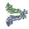
| ||||||||||
|---|---|---|---|---|---|---|---|---|---|---|---|
| 1 | 
| ||||||||||
| Unit cell |
| ||||||||||
| Details | The second dimer is generated by application of the following rotation matrix 1.00x 0.00y 0.00z 207.96 0.00x 1.00y 0.00z 0.7685 0.00x 0.00y -1.00z 131.2386 |
-Components
| #1: Protein | Mass: 50547.785 Da / Num. of mol.: 2 / Mutation: Glu 315 to Glutamine Source method: isolated from a genetically manipulated source Source: (gene. exp.) #2: Chemical | ChemComp-MLT / |   Mass: 134.087 Da / Num. of mol.: 1 / Source method: obtained synthetically / Formula: C4H6O5 Mass: 134.087 Da / Num. of mol.: 1 / Source method: obtained synthetically / Formula: C4H6O5#3: Chemical |   Mass: 192.124 Da / Num. of mol.: 2 / Source method: obtained synthetically / Formula: C6H8O7 Mass: 192.124 Da / Num. of mol.: 2 / Source method: obtained synthetically / Formula: C6H8O7#4: Water | ChemComp-HOH / |  Mass: 18.015 Da / Num. of mol.: 18 / Source method: isolated from a natural source / Formula: H2O Mass: 18.015 Da / Num. of mol.: 18 / Source method: isolated from a natural source / Formula: H2O |
|---|
-Experimental details
-Experiment
| Experiment | Method: X-RAY DIFFRACTION / Number of used crystals: 2 |
|---|
- Sample preparation
| Crystal | Density Matthews: 2.44 Å3/Da / Density % sol: 49.57 % | ||||||||||||||||||||||||||||||||||||
|---|---|---|---|---|---|---|---|---|---|---|---|---|---|---|---|---|---|---|---|---|---|---|---|---|---|---|---|---|---|---|---|---|---|---|---|---|---|
| Crystal grow | Temperature: 291 K / Method: vapor diffusion, hanging drop / pH: 6 Details: PEG 3350, citrate dithiothreitol, pH 6.0, VAPOR DIFFUSION, HANGING DROP at 291K | ||||||||||||||||||||||||||||||||||||
| Crystal grow | *PLUS pH: 5.6 / Method: microdialysis | ||||||||||||||||||||||||||||||||||||
| Components of the solutions | *PLUS
|
-Data collection
| Diffraction | Mean temperature: 298 K |
|---|---|
| Diffraction source | Source: ROTATING ANODE / Type: RIGAKU / Wavelength: 1.5418 Å |
| Detector | Type: XENTRONICS / Detector: AREA DETECTOR / Date: Aug 10, 1998 |
| Radiation | Monochromator: graphite crystal / Protocol: SINGLE WAVELENGTH / Monochromatic (M) / Laue (L): M / Scattering type: x-ray |
| Radiation wavelength | Wavelength: 1.5418 Å / Relative weight: 1 |
| Reflection | Resolution: 2.8→11.5 Å / Num. all: 22978 / Num. obs: 24817 / % possible obs: 92.6 % / Observed criterion σ(F): 2 / Observed criterion σ(I): 1 / Redundancy: 5.8 % / Biso Wilson estimate: 16.05 Å2 / Rmerge(I) obs: 0.1121 / Rsym value: 0.1819 / Net I/σ(I): 5.046 |
| Reflection | *PLUS Highest resolution: 2.5 Å / Lowest resolution: 13 Å / % possible obs: 93.5 % / Rmerge(I) obs: 0.112 |
- Processing
| Software |
| |||||||||||||||||||||||||
|---|---|---|---|---|---|---|---|---|---|---|---|---|---|---|---|---|---|---|---|---|---|---|---|---|---|---|
| Refinement | Method to determine structure: MOLECULAR REPLACEMENT Starting model: PDB Entry 1fuo Resolution: 2.6→8 Å / Cross valid method: THROUGHOUT / σ(F): 1 / Stereochemistry target values: Engh & Huber
| |||||||||||||||||||||||||
| Refinement step | Cycle: LAST / Resolution: 2.6→8 Å
| |||||||||||||||||||||||||
| Refine LS restraints |
| |||||||||||||||||||||||||
| LS refinement shell | Resolution: 2.6→2.71 Å /
| |||||||||||||||||||||||||
| Refinement | *PLUS Highest resolution: 2.6 Å / Rfactor all: 0.179 / Rfactor Rfree: 0.251 / Rfactor Rwork: 0.179 | |||||||||||||||||||||||||
| Solvent computation | *PLUS | |||||||||||||||||||||||||
| Displacement parameters | *PLUS | |||||||||||||||||||||||||
| Refine LS restraints | *PLUS
| |||||||||||||||||||||||||
| LS refinement shell | *PLUS Rfactor Rfree: 0.329 / Rfactor Rwork: 0.235 |
